Understanding Sacroiliac Joint Dysfunction
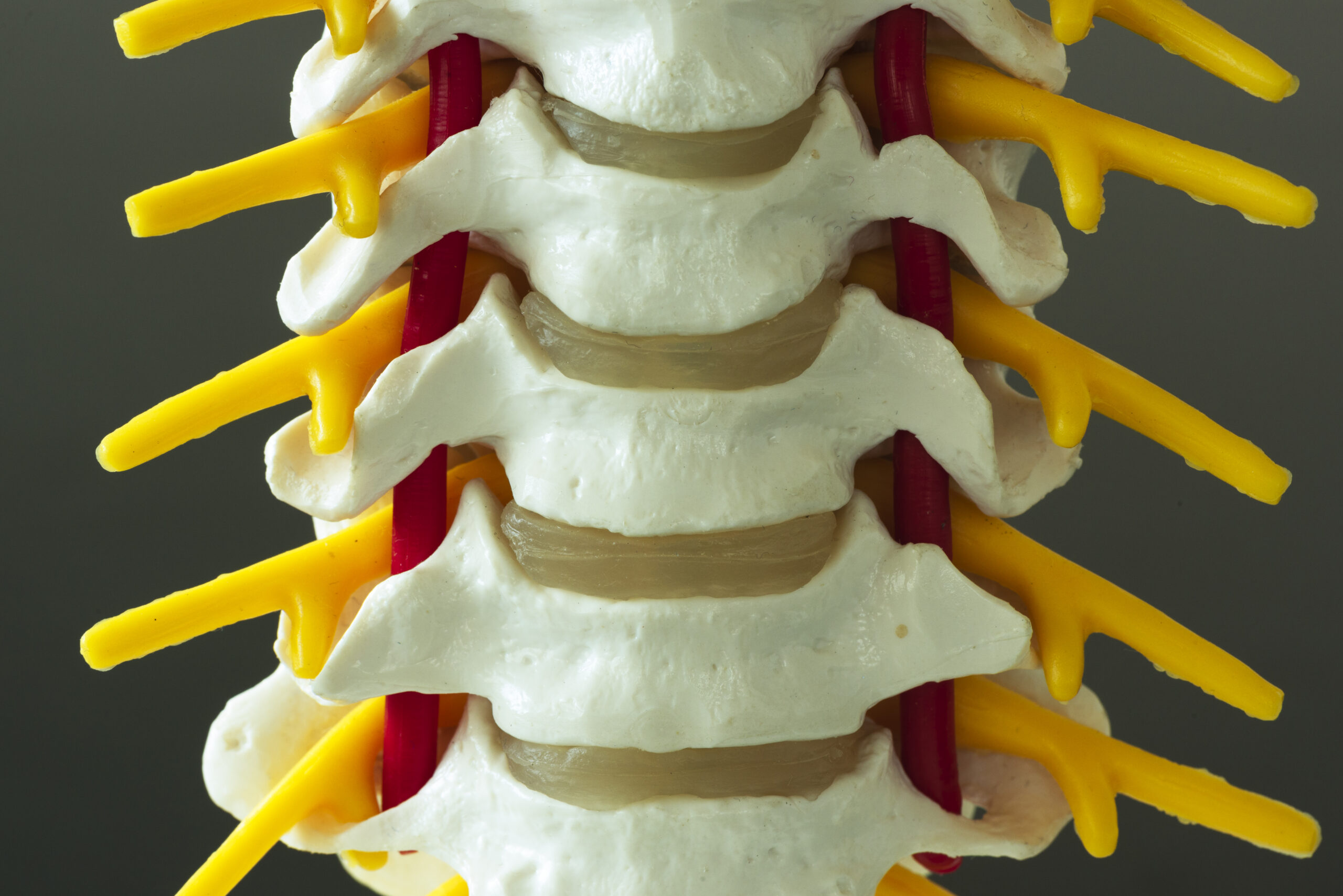
Sacroiliac (SI) joint dysfunction occurs when the joints connecting the spine to the pelvis become misaligned or inflamed, causing pain in the lower back, hips, or legs. This condition can mimic other spinal issues, making accurate diagnosis critical. Common symptoms include localized tenderness, radiating discomfort, and difficulty with movements like standing or climbing stairs. While SI joint dysfunction is often linked to biomechanical imbalances, trauma, or pregnancy, its overlap with conditions like lumbar disc herniation or sciatica requires precise evaluation. Chiropractors play a key role in identifying this issue through targeted assessments, ensuring patients receive appropriate care. Without proper diagnosis, treatments may fail to address the root cause, leading to prolonged discomfort. Understanding the diagnostic tools available helps both practitioners and patients navigate effective management strategies.
Clinical Evaluation Techniques
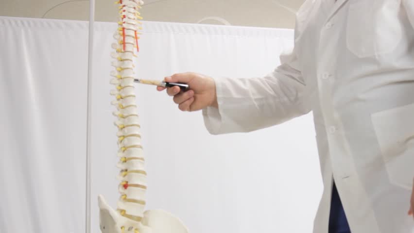
Chiropractors begin by conducting a thorough physical examination to assess SI joint function. This includes palpation to detect tenderness or asymmetry, as well as range-of-motion tests to identify movement restrictions. Specific orthopedic tests, such as the FABER (Flexion, Abduction, External Rotation) maneuver or the Gaenslen’s test, are commonly used to provoke or alleviate pain, helping confirm SI joint involvement. These manual techniques rely on the practitioner’s expertise to differentiate SI joint dysfunction from other sources of back pain. While subjective, they provide valuable baseline information. Patients are often asked to describe pain patterns, which further guides the evaluation. These initial steps are essential for narrowing down potential causes and determining whether advanced diagnostic tools are needed for a definitive diagnosis.
Imaging and Diagnostic Tools
Imaging studies are frequently used to support clinical findings in diagnosing SI joint dysfunction. X-rays can reveal structural abnormalities, such as arthritis or misalignment, though they may not always capture soft tissue issues. Magnetic resonance imaging (MRI) offers detailed views of ligaments, discs, and surrounding tissues, making it useful for identifying inflammation or damage not visible on X-rays. Computed tomography (CT) scans may be employed for clearer bone imaging in complex cases. Injections, like diagnostic SI joint blocks, involve administering anesthetic to determine if the joint is the pain source. While these tools enhance accuracy, they are typically used alongside clinical exams. Chiropractors interpret results in context, avoiding over-reliance on imaging alone. The choice of tool depends on symptom severity and the need for further investigation.
Functional Assessments and Gait Analysis
Functional assessments evaluate how the SI joint interacts with movement patterns. Chiropractors observe patients during activities like walking, squatting, or standing to identify compensatory mechanics that may contribute to dysfunction. Gait analysis, often using video or specialized software, highlights irregularities in weight distribution or pelvic motion. These assessments help pinpoint biomechanical imbalances, such as leg length discrepancies or muscle weakness, that could exacerbate SI joint strain. By integrating functional data with manual exams, chiropractors gain a holistic view of the patient’s condition. This approach is particularly useful for athletes or individuals with repetitive movement patterns. Functional evaluations also guide personalized treatment plans, ensuring interventions address both symptoms and underlying causes.
Integrating Diagnostic Findings for Treatment
Once diagnostic tools confirm SI joint dysfunction, chiropractors develop tailored treatment strategies. Manual adjustments, soft tissue therapies, and rehabilitative exercises are commonly employed to restore joint mobility and reduce inflammation. The choice of intervention depends on the severity and underlying factors identified during evaluation. For example, a patient with significant instability may benefit from stabilization exercises, while someone with acute inflammation might require anti-inflammatory modalities. Chiropractors also consider patient preferences and lifestyle when designing care plans. Regular reassessments ensure treatments remain effective and adaptable. By aligning interventions with diagnostic insights, practitioners enhance outcomes and promote long-term musculoskeletal health. Collaboration with other healthcare providers may also be necessary for comprehensive care.
Chiropractic Role in Ongoing Management
Chiropractors emphasize ongoing monitoring to manage SI joint dysfunction effectively. Regular follow-ups allow for tracking progress and adjusting treatment as needed. Patients are often educated on self-care techniques, such as posture correction or low-impact exercises, to prevent recurrence. Chiropractors may also recommend ergonomic modifications or assistive devices based on diagnostic findings. For chronic cases, a multidisciplinary approach involving physical therapy or pain management specialists might be advised. The goal is to empower patients with knowledge and tools for long-term wellness. By combining diagnostic precision with patient-centered care, chiropractors help individuals achieve sustained relief and improved quality of life.