Understanding the Role of Palpation in Diagnosis
Accurate diagnosis of musculoskeletal conditions relies heavily on clinical examination, with palpation serving as a critical tool for distinguishing sacroiliac joint dysfunction from lumbar spine issues. Both conditions can present with lower back pain, but their underlying causes and affected structures differ. Palpation allows clinicians to identify specific tenderness, asymmetry, or abnormal movement patterns that may point to the source of discomfort. By systematically evaluating key anatomical landmarks, practitioners can gather valuable information to guide further assessment. However, it is essential to recognize that palpation alone is not definitive and should be combined with other diagnostic methods. This approach ensures a comprehensive evaluation, reducing the risk of misdiagnosis. Patients should always consult a qualified healthcare provider for personalized assessments, as individual variations can influence findings.
Palpation Techniques for the Sacroiliac Joint
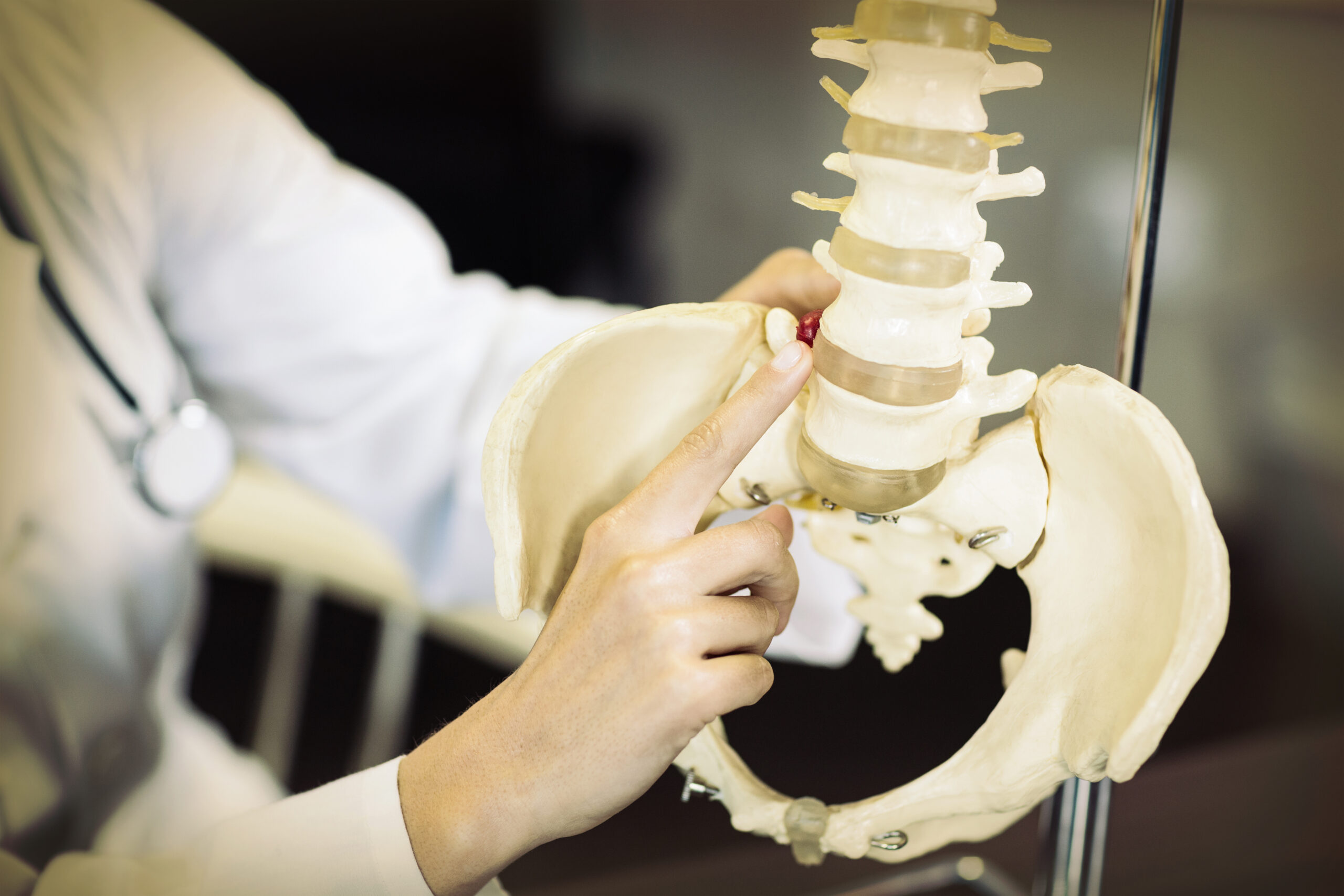
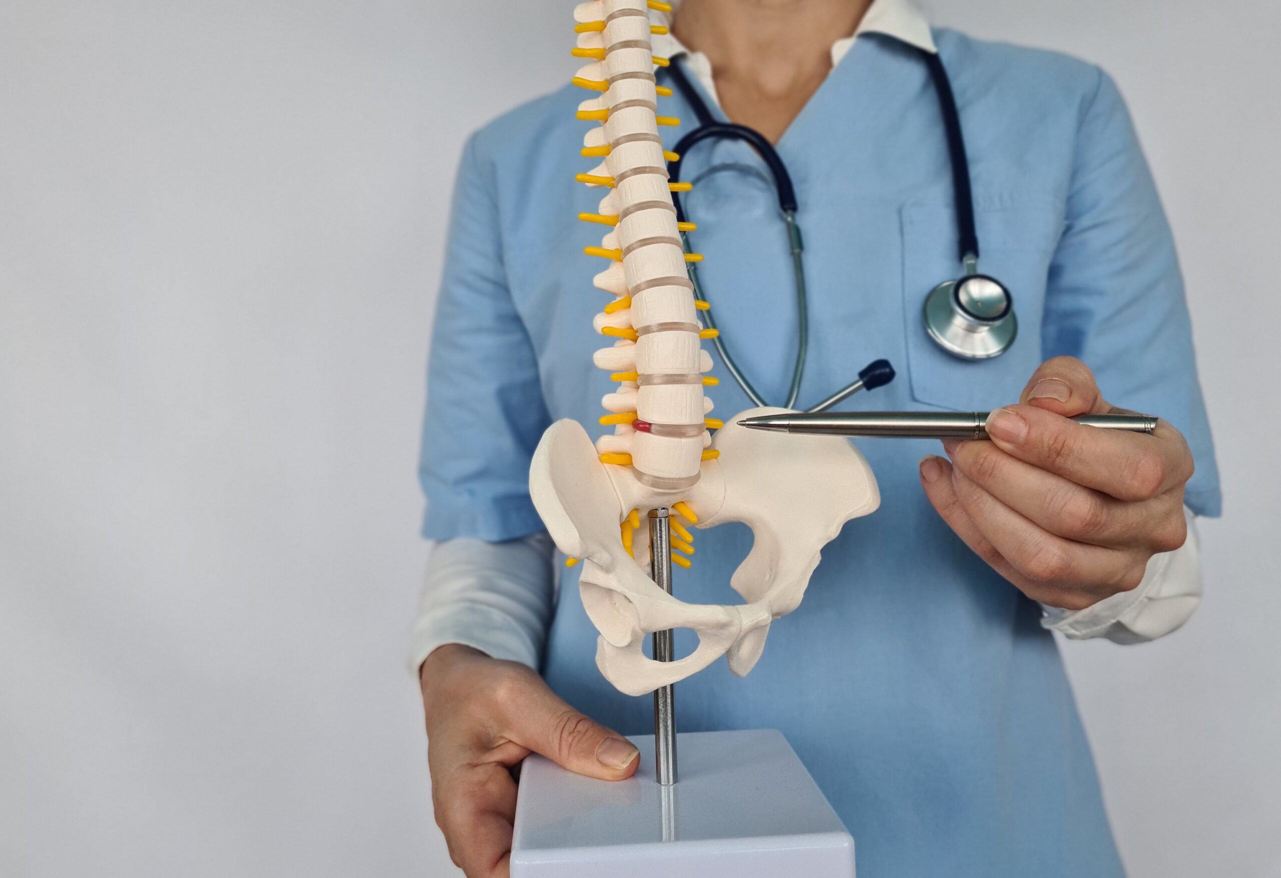
Palpation of the sacroiliac (SI) joint involves assessing the posterior superior iliac spines (PSIS) and the sacral sulci. Clinicians typically apply gentle pressure to these areas to detect tenderness, which is common in SI joint dysfunction. A key differentiator is the presence of pain localized to the SI joint region, often radiating to the gluteal area or posterior thigh. Asymmetry in the PSIS or a palpable gap between the sacrum and ilium may also indicate dysfunction. Additionally, the presence of a positive Faber test (Patrick’s test) can support SI joint involvement. However, these findings should be interpreted alongside other clinical signs. The goal is to isolate the SI joint’s contribution to pain, avoiding confusion with lumbar spine issues that may mimic similar symptoms. Proper technique and patient positioning are crucial for reliable results.
Evaluating the Lumbar Spine Through Palpation
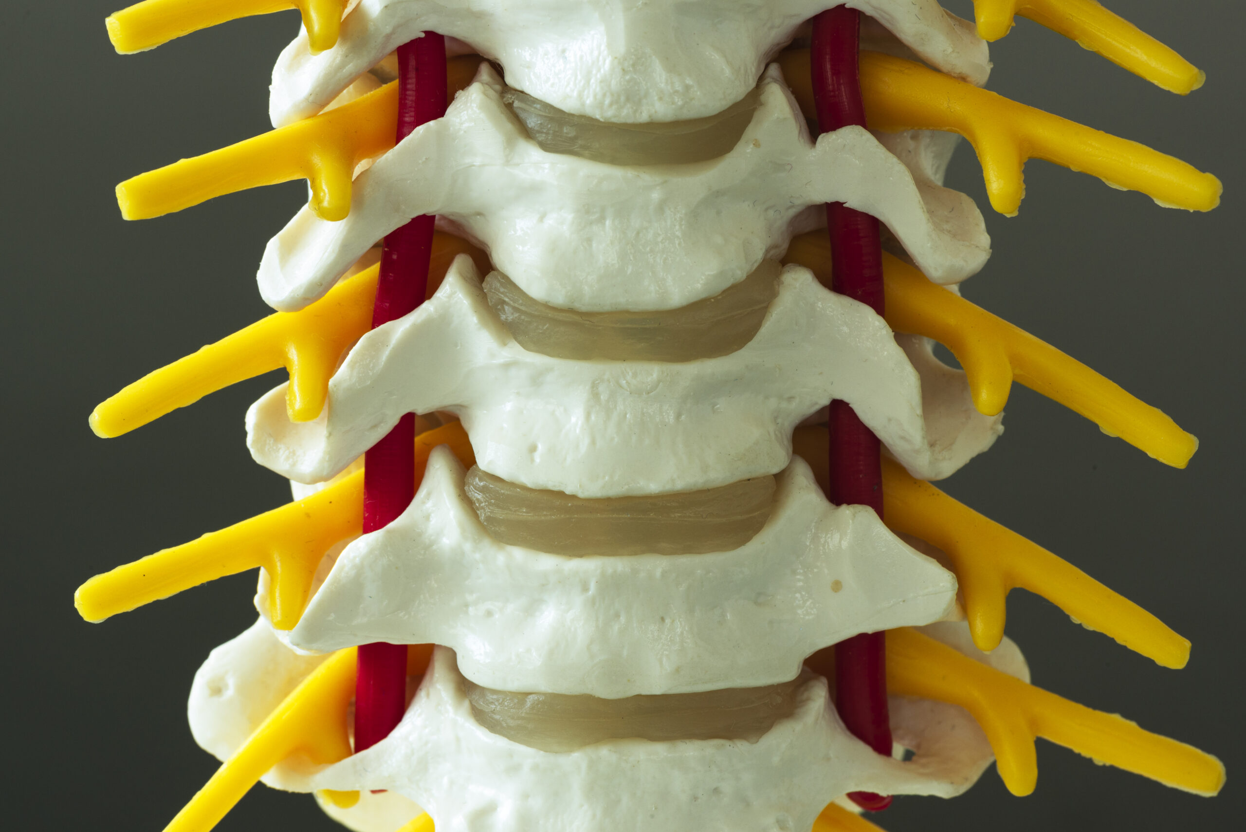
When assessing the lumbar spine, palpation focuses on the spinous processes, transverse processes, and paraspinal muscles. Tenderness along the midline or lateral to the spine may suggest lumbar disc herniation, facet joint irritation, or muscle strain. Clinicians often check for tenderness in the lumbar erector spinae and assess for guarding or spasm, which can indicate nerve root involvement. Unlike SI joint dysfunction, lumbar spine issues typically present with pain that radiates into the lower extremities, following a dermatomal pattern. Palpation may also reveal restricted range of motion or tenderness upon axial loading. It is important to differentiate these findings from those associated with SI joint dysfunction, as treatment approaches vary significantly. Combining palpation with neurological and orthopedic tests enhances diagnostic accuracy, ensuring targeted interventions for the patient’s specific condition.
Identifying Key Differentiation Points
Several factors help distinguish SI joint dysfunction from lumbar spine issues during palpation. SI joint pain is often deep, localized to the gluteal region, and exacerbated by weight-bearing activities, while lumbar spine pain tends to be more diffuse and worsened by forward bending. Palpation of the SI joint may elicit pain when applying pressure to the PSIS or sacral crests, whereas lumbar spine issues typically cause tenderness along the spinous processes. Additionally, SI joint dysfunction may present with a positive Gaenslen’s test, where pain is reproduced by lying on the unaffected side and extending the affected leg. In contrast, lumbar spine conditions may show pain with straight leg raise tests. Recognizing these distinctions is vital for accurate diagnosis, as misidentification can lead to ineffective treatment. A thorough, systematic approach ensures that both conditions are properly evaluated and managed.
The Importance of Clinical Correlation
Palpation findings must be interpreted within the broader context of the patient’s history and physical examination. For example, a patient with a history of trauma may present with SI joint instability, while degenerative changes in the lumbar spine could explain chronic pain. Clinicians should consider associated symptoms such as radiculopathy, neurological deficits, or referred pain patterns. Combining palpation with range-of-motion testing, neurological assessments, and imaging (when indicated) provides a more complete picture. It is also crucial to rule out systemic conditions or other pathologies that may mimic SI joint or lumbar spine issues. By integrating multiple diagnostic tools, practitioners can minimize errors and develop effective treatment plans. Patients should be encouraged to communicate their symptoms openly to facilitate accurate evaluation and care.
Conclusion: Collaborating for Accurate Diagnosis
Palpation techniques play a vital role in differentiating sacroiliac joint dysfunction from lumbar spine issues, but they require skill and clinical judgment. While specific findings can guide the diagnostic process, they should not be viewed in isolation. A comprehensive approach that includes patient history, physical examination, and, when necessary, imaging ensures a reliable diagnosis. Chiropractors and other healthcare providers must stay current with evidence-based practices to enhance their diagnostic accuracy. Patients benefit from this thorough evaluation, as it leads to targeted interventions and improved outcomes. Ultimately, collaboration between clinicians and patients fosters trust and ensures that care is tailored to individual needs. By prioritizing accurate diagnosis, healthcare providers can address the root cause of discomfort and promote long-term wellness.