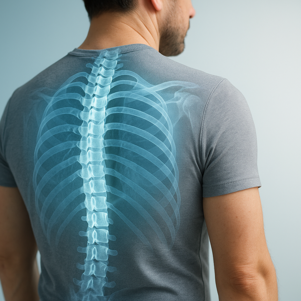
Understanding the Thoracic Spine: An Overview
The human spine is one of the most complex and essential structures in the body, providing support, flexibility, and protection for the spinal cord. Among its three primary regions, the thoracic spine stands out as the longest and most structurally unique segment. Whether you are a medical professional, a student of anatomy, or simply someone curious about how the body works, understanding the thoracic spine is fundamental to appreciating how the upper body functions as a whole.
What Is the Thoracic Spine?
The thoracic spine refers to the middle section of the vertebral column, situated between the cervical spine (the neck region) above and the lumbar spine (the lower back) below. It is commonly referred to as the mid back or upper back spine. This region plays a critical role in maintaining posture, anchoring the rib cage, and protecting the vital organs housed within the chest cavity.
In thoracic spine anatomy, this segment is distinguished by its unique attachment to the ribs, which sets it apart from every other region of the spine. This characteristic gives the thoracic spine remarkable stability but comparatively limited range of motion when contrasted with the cervical or lumbar regions.
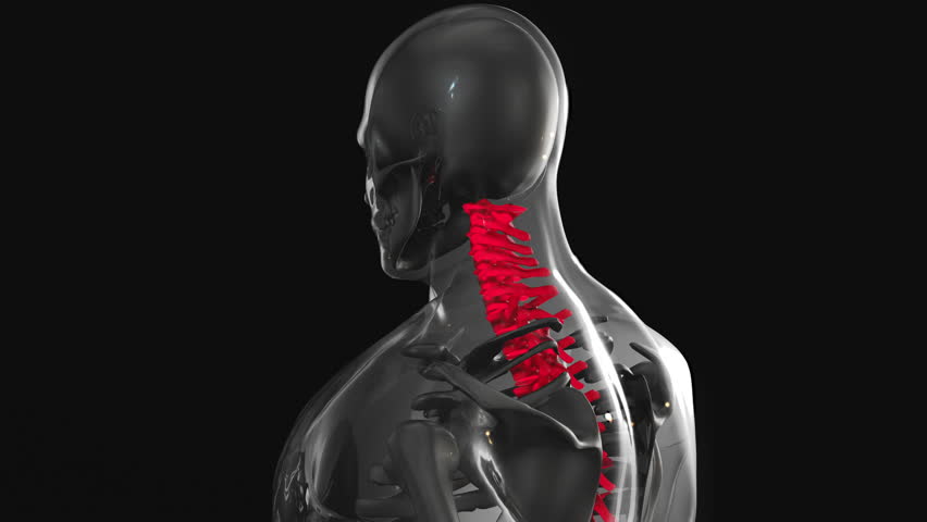
The Vertebrae of the Thoracic Spine: T1 Through T12
The thoracic spine is composed of twelve vertebrae, labeled T1 through T12. Each vertebra is numbered sequentially from top to bottom, with T1 being the uppermost and T12 being the lowest. These mid back vertebrae are progressively larger in size as they descend, a design that reflects the increasing mechanical load each vertebra must bear.
Here is a closer look at the individual segments within the thoracic spine:
- T1 – T4 (Upper Thoracic): These vertebrae are situated near the base of the neck and top of the upper back spine. They are closely associated with the upper ribs and contribute to movements of the shoulders and arms through their proximity to the brachial plexus nerve network.
- T5 – T8 (Middle Thoracic): This central group of vertebrae forms the core of the thoracic region. They provide the backbone for the mid-section of the rib cage and play a primary role in trunk stability and respiratory mechanics.
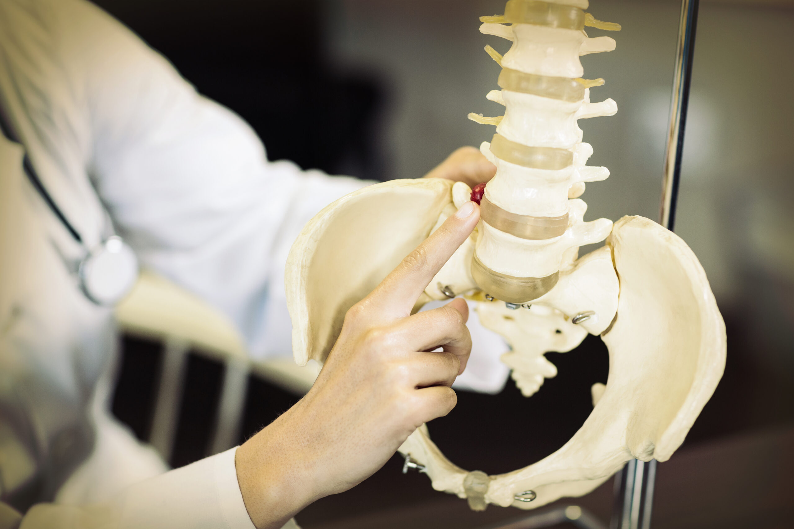
- T9 – T12 (Lower Thoracic): The lower thoracic vertebrae transition toward the lumbar region. T12, in particular, is a transitional vertebra that shares characteristics of both thoracic and lumbar vertebrae. These lower segments support greater weight and are involved in a wider range of motion than the upper thoracic vertebrae.
Key Anatomical Features of the Thoracic Vertebrae
Each thoracic vertebra shares several defining anatomical characteristics that are central to thoracic spine anatomy. Understanding these features helps explain how the thoracic spine performs its many functions.
The Vertebral Body
The vertebral body is the large, cylindrical anterior portion of each vertebra. In the thoracic spine, these bodies are heart-shaped when viewed from above and gradually increase in size from T1 to T12. The vertebral bodies bear the compressive forces of body weight and transmit them downward through the spine.
The Spinous Process
One of the most recognizable features of thoracic vertebrae is their long, downward-angled spinous processes. These bony projections extend posteriorly and inferiorly, giving the thoracic spine its characteristic steep slope when palpated along the back. The spinous processes serve as attachment points for muscles and ligaments that help stabilize the spine.
Costal Facets (Rib Articulations)
Perhaps the most defining anatomical feature of the mid back vertebrae is the presence of costal facets — specialized joint surfaces that articulate with the ribs. Each thoracic vertebra typically has two pairs of costal facets: demifacets located on the vertebral body and transverse costal facets on the transverse processes. These articulations form the costovertebral joints, which are essential to respiratory function.
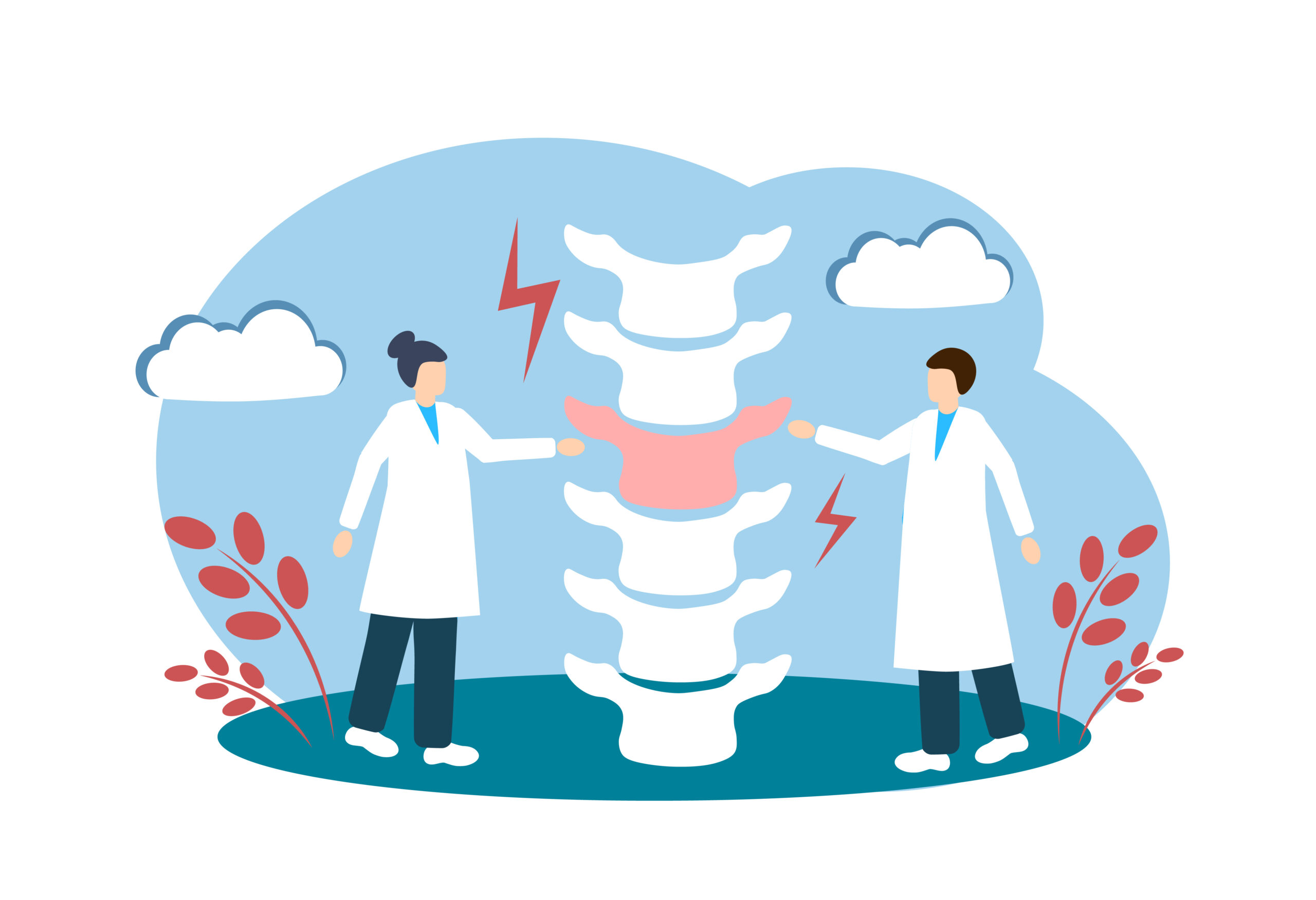
The Intervertebral Discs
Between each pair of vertebrae lies an intervertebral disc, composed of a tough outer ring called the annulus fibrosus and a soft, gel-like center called the nucleus pulposus. In the thoracic region, these discs are relatively thin compared to those in the lumbar spine, contributing to the reduced range of motion in this area.
The Spinal Canal
Running through the vertebral arch of each vertebra is the spinal canal, through which the spinal cord passes. The thoracic spinal canal is notably narrower than those in the cervical and lumbar regions, which means that any injury, disc herniation, or structural abnormality in this area carries a higher risk of spinal cord compression.
The Role of the Thoracic Spine in the Body
The thoracic spine serves several essential physiological and structural functions. Its design reflects the unique demands placed upon it by the human body.
Structural Support and Posture
The thoracic spine forms the foundation of the upper back spine and provides the structural framework necessary for an upright posture. Its natural kyphotic curve — a gentle, outward curvature when viewed from the side — complements the inward curves of the cervical and lumbar regions, creating the characteristic S-shape of the entire spinal column. This curvature is critical for evenly distributing mechanical loads across the spine.
Protection of Internal Organs
The rib cage, which attaches directly to the thoracic vertebrae via the costovertebral joints, forms a protective bony enclosure around the heart, lungs, and major blood vessels. By anchoring the ribs, the thoracic spine plays an indirect but vital role in safeguarding these essential organs from injury.