Understanding the Vertebra: The Building Block of Your Spine
The human spine is one of the most remarkable structures in the body, providing both stability and flexibility while protecting the delicate spinal cord within. At the core of this complex system lies a single, fundamental unit — the vertebra. Understanding vertebra anatomy is essential not only for medical professionals but also for anyone seeking to understand how chiropractic care works and why spinal health matters so deeply to overall well-being.
What Exactly Is a Vertebra?
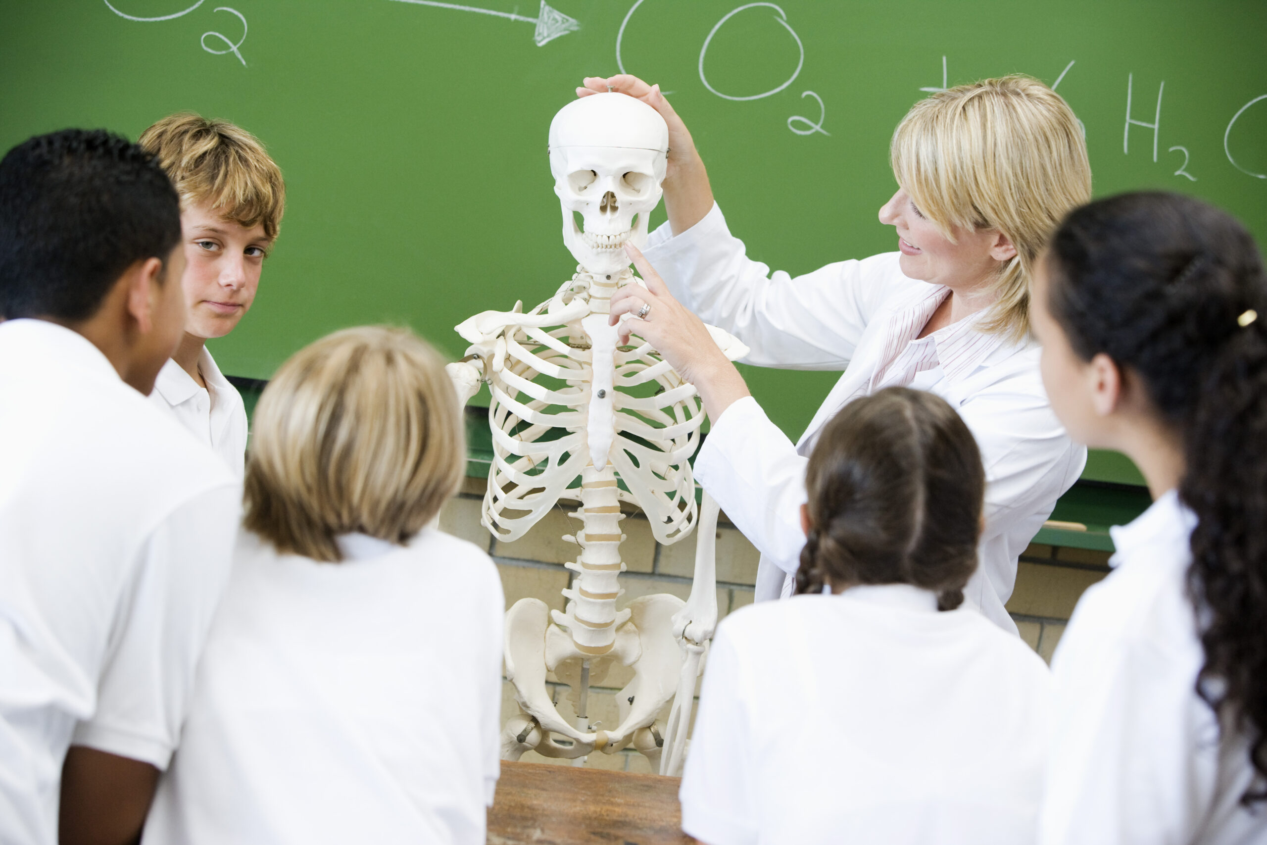
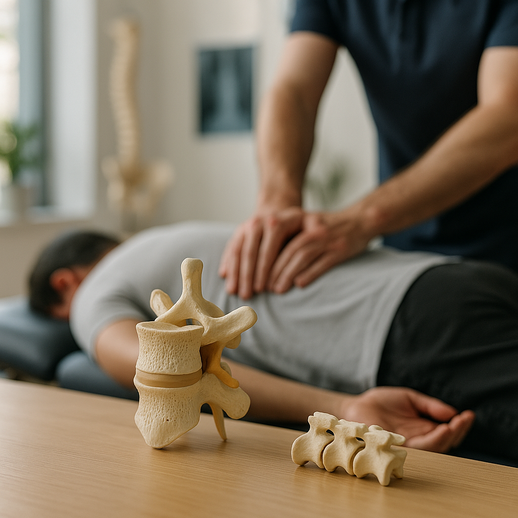
A vertebra is an individual spine bone that forms part of the vertebral column, commonly known as the backbone or spine. Each vertebra is a uniquely shaped bone designed to perform several critical functions simultaneously: bearing the weight of the body above it, protecting the spinal cord, and allowing a controlled range of motion. The plural form of this term is vertebrae, and in a healthy adult human, there are typically 33 of these individual spine bones stacked in a carefully organized column.
While each vertebra shares a common basic structure, the specific shape and size vary depending on its location in the spine and the functional demands placed upon it. Understanding these differences helps explain why certain regions of the spine are more susceptible to injury, degeneration, or misalignment — all of which are central concerns in chiropractic practice.
The Anatomy of a Single Vertebra
Each spinal vertebra is composed of several distinct anatomical components, each serving a specific and vital purpose. The key structures include:
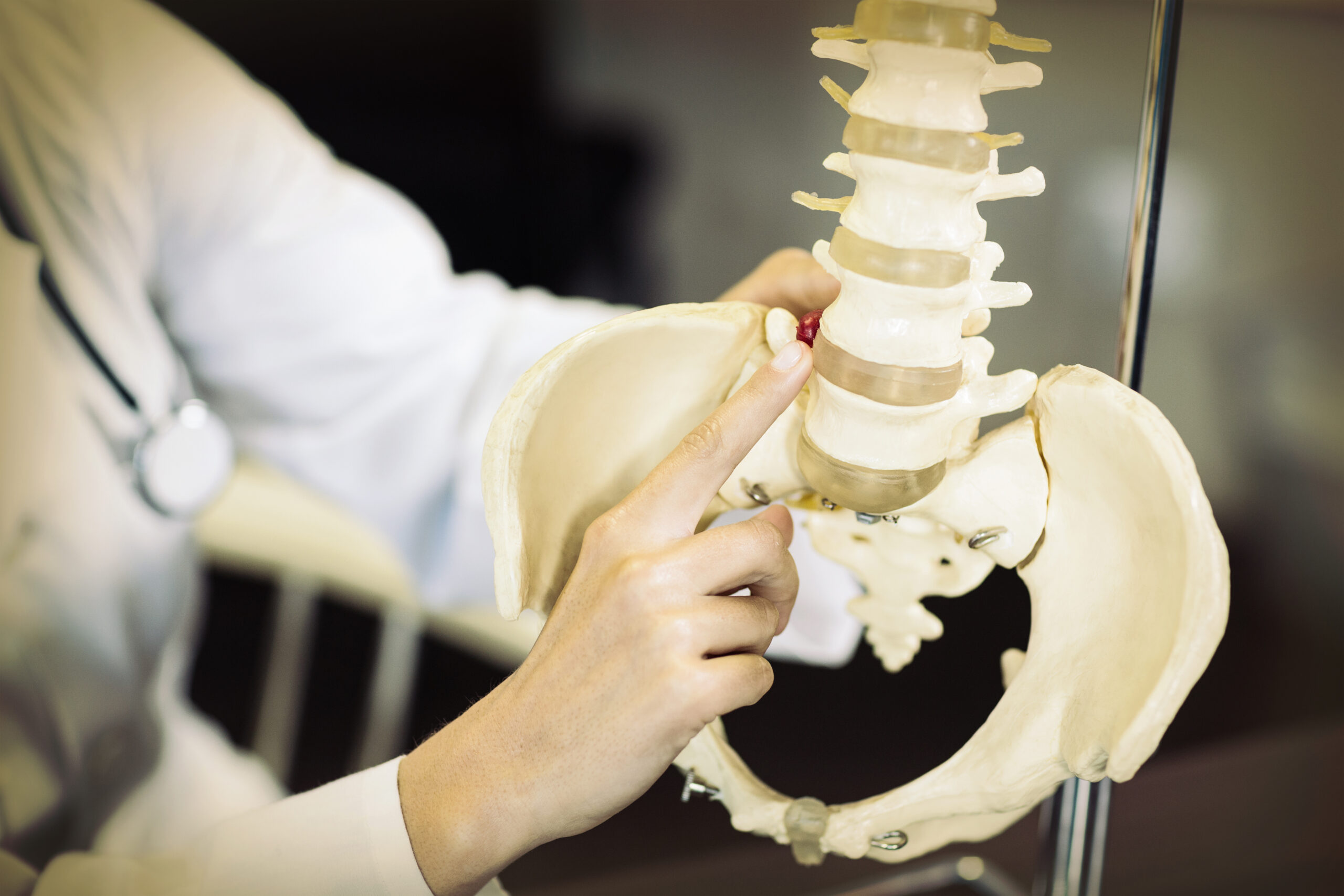
- The Vertebral Body: The vertebral body is the large, cylindrical front portion of the vertebra. It is the primary weight-bearing component and is made of dense, compact bone on the outside with a spongy interior. The vertebral bodies of adjacent vertebrae are separated by intervertebral discs, which act as cushions and shock absorbers.
- The Vertebral Arch: Extending from the back of the vertebral body, the vertebral arch forms a ring-like structure that, together with the vertebral body, creates the vertebral foramen — the opening through which the spinal cord passes.
- The Spinous Process: This is the bony projection that you can feel along the center of your back. It serves as an attachment point for muscles and ligaments.
- The Transverse Processes: These are two lateral projections extending from either side of the vertebral arch, also serving as muscle and ligament attachment points.
- The Articular Processes: There are four articular processes per vertebra — two superior and two inferior — which form the facet joints that connect adjacent vertebrae to one another, enabling controlled movement while maintaining stability.
- The Pedicles and Laminae: These are the connecting portions of the vertebral arch. The pedicles connect the arch to the vertebral body, while the laminae form the posterior part of the arch.
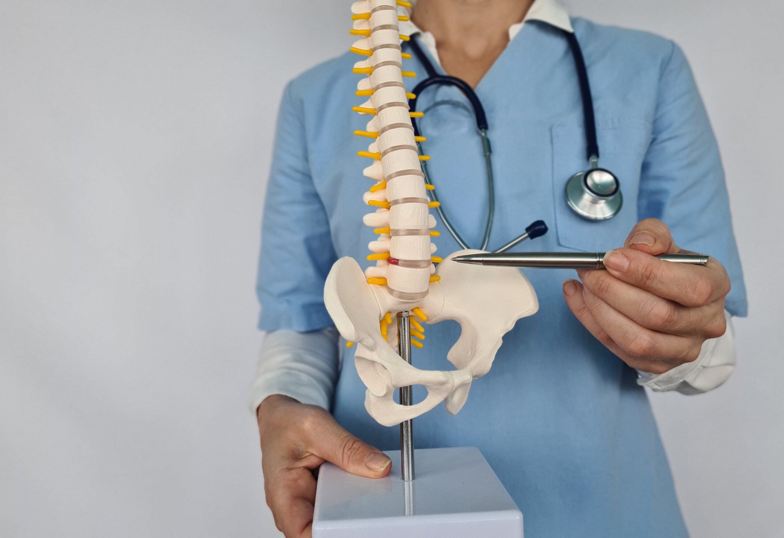
The Five Regions of the Spinal Column
To appreciate how a spinal vertebra chiropractic assessment is conducted, it helps to understand how the vertebral column is organized into five distinct regions, each with its own characteristics and range of motion:
- Cervical Region (C1–C7): The seven vertebrae of the neck form the cervical spine. These are the smallest and most delicate vertebrae, designed to support the weight of the head and allow the greatest range of motion. The top two — the atlas (C1) and the axis (C2) — have unique shapes that allow the head to nod and rotate.
- Thoracic Region (T1–T12): The twelve thoracic vertebrae form the mid-back and articulate with the ribs, creating the thoracic cage. They are larger than cervical vertebrae but offer a more restricted range of motion due to their attachment to the rib cage.
- Lumbar Region (L1–L5): The five lumbar vertebrae are the largest and most robust of the movable vertebrae. They bear the greatest proportion of body weight and are frequently the site of injury, herniation, and chiropractic intervention.
- Sacral Region (S1–S5): These five vertebrae are fused in adults to form the sacrum, a triangular bone that connects the spine to the pelvis.
- Coccygeal Region: The remaining three to five small bones fuse to form the coccyx, commonly known as the tailbone.
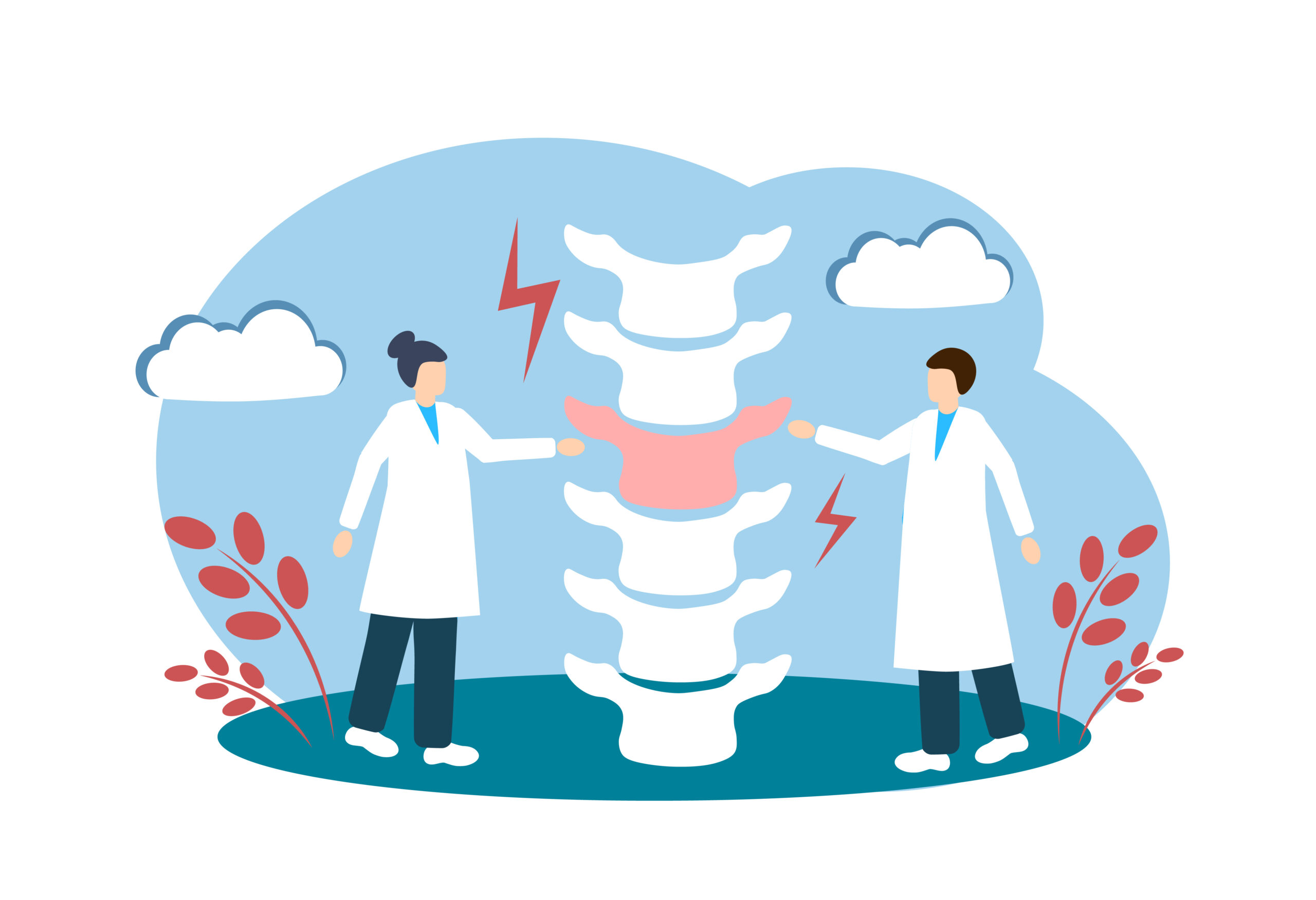
The Role of Intervertebral Discs
Between most vertebral bodies lie intervertebral discs — fibrocartilaginous structures composed of a tough outer ring called the annulus fibrosus and a gel-like center called the nucleus pulposus. These discs serve as the spine’s shock absorbers, distributing compressive forces evenly across the vertebral bodies during movement and weight-bearing activities.
When these discs become damaged, dehydrated, or displaced — conditions commonly referred to as disc herniation or disc degeneration — they can press upon adjacent nerve roots, causing pain, numbness, or weakness that may radiate into the limbs. Addressing the surrounding vertebral structures is a primary focus of many chiropractic treatment approaches.
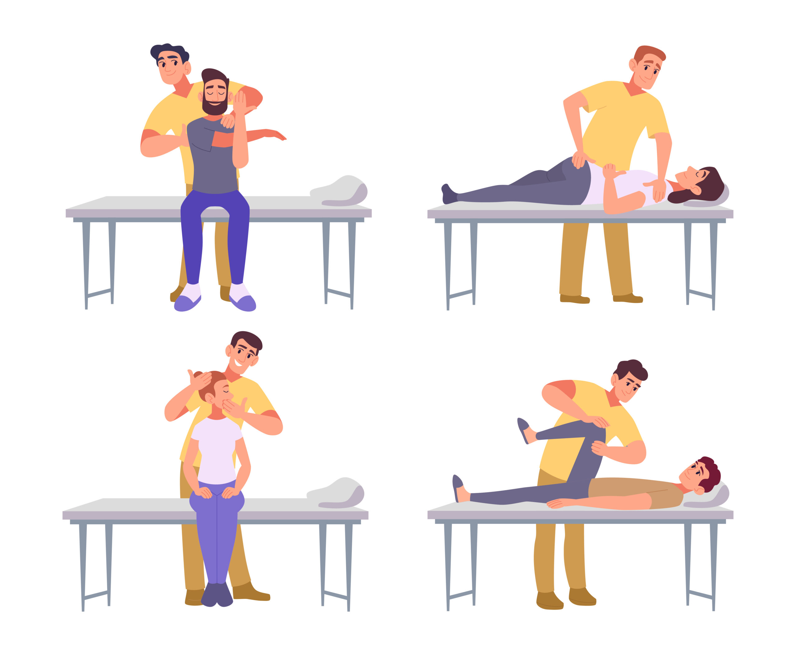
How Vertebra Anatomy Relates to Chiropractic Care
Chiropractic care is a regulated healthcare discipline that focuses on the diagnosis, treatment, and prevention of disorders related to the musculoskeletal system — particularly the spine. The relationship between vertebra anatomy and chiropractic practice is both foundational and inseparable.
Chiropractors are specifically trained to identify and address vertebral subluxations — a term used within the chiropractic profession to describe situations in which one or more vertebrae are positioned or moving in a manner that may interfere with normal nerve function or create undue stress on surrounding tissues.