Understanding the Sacrum in Chiropractic Anatomy
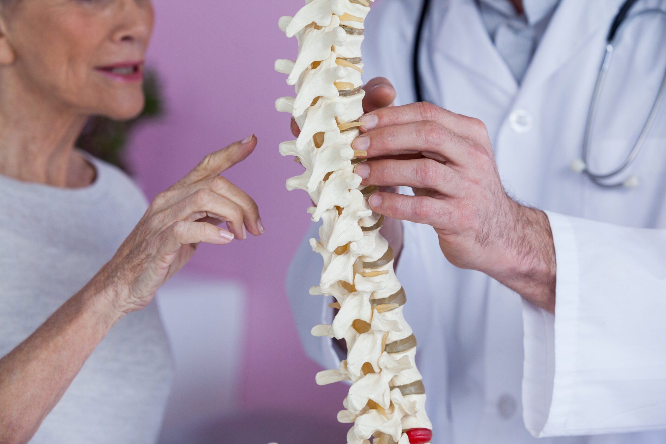
The sacrum is one of the most important structural components of the human spine and pelvis. In chiropractic practice, a thorough understanding of sacrum anatomy is essential for diagnosing and treating a wide range of musculoskeletal conditions. Whether you are a patient seeking answers about lower back pain or simply curious about how your body is structured, learning about the sacral spine can provide valuable insight into how your body moves, balances, and functions as a whole.
What Is the Sacrum?
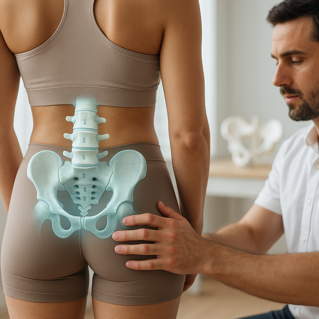
The sacrum is a large, triangular bone located at the base of the spine, just below the lumbar vertebrae and above the coccyx, commonly known as the tailbone. This triangular bone sits at the very center of the pelvis, forming the posterior wall of the pelvic girdle. It serves as the keystone of the skeletal structure, connecting the spinal column to the hip bones on either side.
In terms of sacrum anatomy, the bone is formed by the fusion of five individual sacral vertebrae, typically labeled S1 through S5. This fusion process begins during adolescence and is generally complete by the time a person reaches their late twenties. The result is a solid, wedge-shaped bone that is broader at the top and narrows toward the bottom, giving it its characteristic triangular appearance.
Key Anatomical Features of the Sacral Spine
The sacral spine is composed of several important anatomical structures that chiropractors closely examine during assessment and treatment. Understanding these features helps explain why the sacrum plays such a critical role in overall spinal health.
- Sacral Foramina: These are small openings on both the anterior and posterior surfaces of the sacrum through which sacral nerve roots pass. There are four pairs of anterior sacral foramina and four pairs of posterior sacral foramina, allowing nerves to travel to the pelvis, legs, and lower organs.
- Sacral Canal: A continuation of the spinal canal, the sacral canal runs through the interior of the sacrum and houses the cauda equina, which is the bundle of nerve roots at the lower end of the spinal cord.
- Sacral Promontory: This is the anterior projection at the superior edge of the sacrum, which forms an important anatomical landmark in the pelvis.
- Auricular Surface: Located on the lateral sides of the sacrum, the auricular surfaces articulate with the iliac bones of the pelvis, forming the sacroiliac joints.
- Sacral Crest: A series of ridges along the posterior surface of the sacrum, which serve as attachment points for ligaments and muscles.
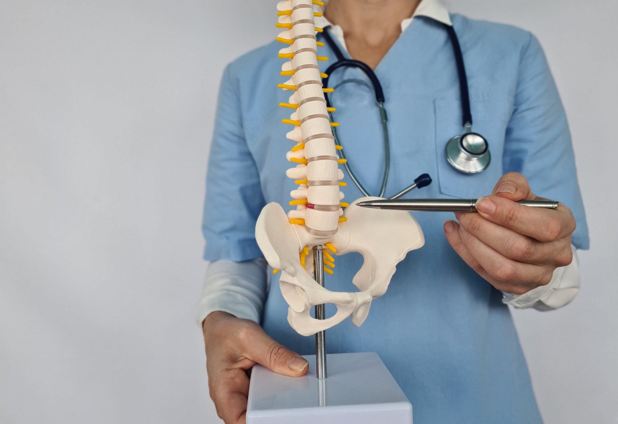
The Role of the Sacrum in Pelvic Structure
The triangular bone of the pelvis serves as far more than just a structural anchor. Its position between the two iliac bones of the hip means that it must withstand and distribute significant mechanical forces that travel down through the spine and up through the legs. The sacrum essentially acts as a bridge between the axial skeleton, which includes the skull, vertebral column, and rib cage, and the appendicular skeleton, which includes the limbs.
The sacroiliac joints, which connect the sacrum to the pelvis on each side, are particularly important in chiropractic care. These joints are supported by some of the strongest ligaments in the human body and allow for a limited but crucial range of movement. When these joints become misaligned, inflamed, or restricted, patients often experience pain in the lower back, buttocks, hips, and even down the legs.
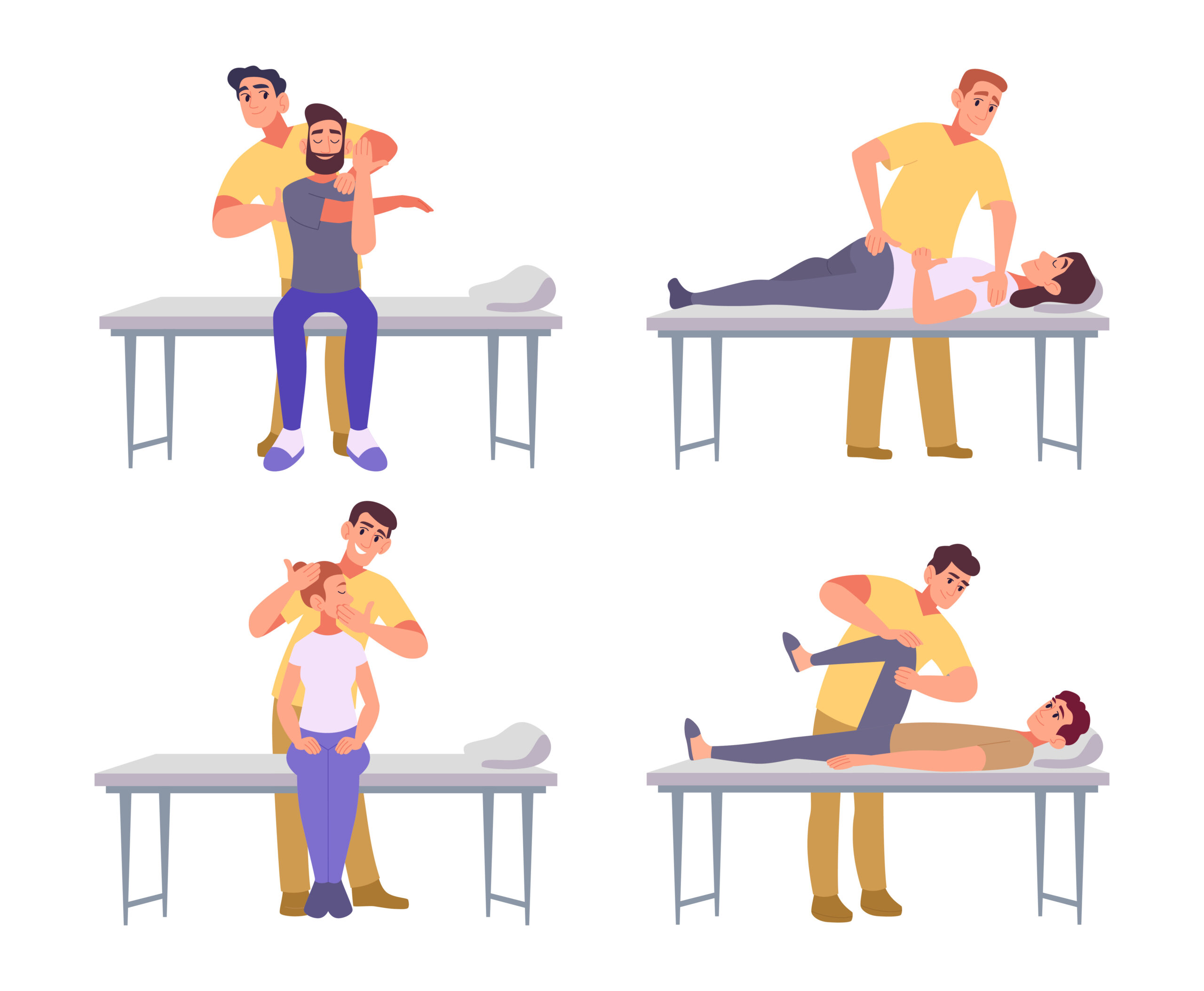
The Sacrum in Chiropractic Practice
In sacrum chiropractic care, this bone is a primary focus of assessment and treatment. Chiropractors are trained to identify misalignments, known as subluxations, within the sacral region and to apply precise adjustments that restore proper alignment and function. Because the sacrum is so central to the structure of the spine and pelvis, even minor misalignments in this area can have wide-reaching effects on the rest of the body.
Patients who present with the following conditions may benefit from chiropractic evaluation of the sacral region:
- Chronic lower back pain
- Sacroiliac joint dysfunction
- Sciatica or radiating leg pain
- Hip pain and stiffness
- Pelvic imbalance or postural issues
- Pain during pregnancy or postpartum discomfort
- Tailbone pain following trauma or injury
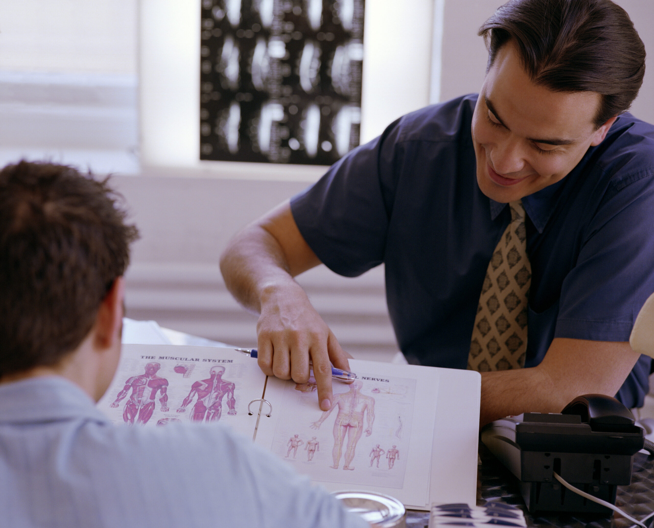
During a chiropractic evaluation, the practitioner will typically assess the position and mobility of the sacrum through both physical palpation and postural analysis. Imaging studies such as X-rays may also be used to gain a more detailed view of the sacral spine and its relationship to the surrounding structures.
Sacral Motion and Its Importance
One of the unique aspects of sacrum anatomy that chiropractors pay close attention to is the concept of sacral motion. Although the sacrum appears as a fixed structure, it is actually capable of subtle movements that are important for spinal health. Two primary types of sacral movement are commonly discussed in chiropractic literature:
- Nutation: This refers to the forward tilting of the sacrum relative to the ilium. During nutation, the sacral promontory moves anteriorly and inferiorly, while the apex of the sacrum moves posteriorly. This movement helps to stabilize the pelvis and is typically associated with activities such as standing and walking.
- Counternutation: This is the opposite movement, where the sacral promontory tilts posteriorly and superiorly. Counternutation is often associated with forward bending and flexion of the lumbar spine.
Additional Resources