Understanding the Lumbar Spine: A Complete Guide to Your Lower Back
The lumbar spine is one of the most critical regions of the human body, yet it remains poorly understood by many people — until pain or injury brings it to their attention. Whether you are a healthcare professional, a patient recovering from a back injury, or simply someone curious about human anatomy, understanding the lumbar spine can provide valuable insight into how your body moves, supports itself, and functions every day.
What Is the Lumbar Spine?
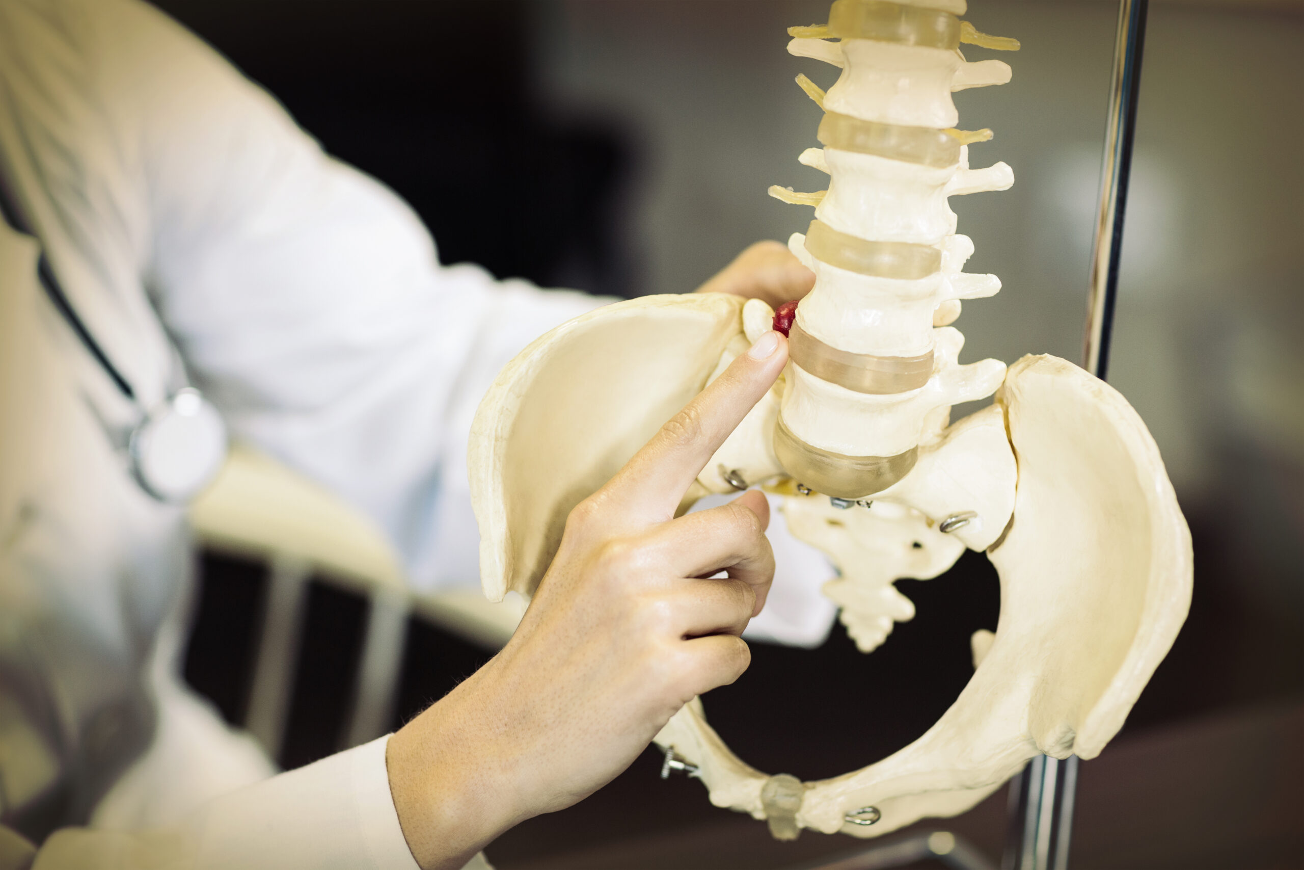
The lumbar spine refers to the lower portion of the vertebral column, situated between the thoracic spine above and the sacrum below. In lumbar spine anatomy, this region is characterized by five large vertebrae that bear the majority of the body’s weight and facilitate a wide range of motion. The lower spine lumbar region plays a fundamental role in nearly every physical activity a person performs, from sitting and standing to bending and lifting.
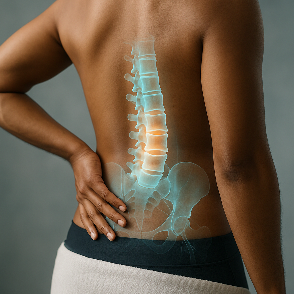
Because this section of the spine endures considerable mechanical stress throughout a person’s lifetime, it is also one of the most common sites of pain, injury, and degenerative conditions. Understanding its structure is the first step toward appreciating why it is so vulnerable — and so vital.
Lumbar Spine Anatomy: The Building Blocks
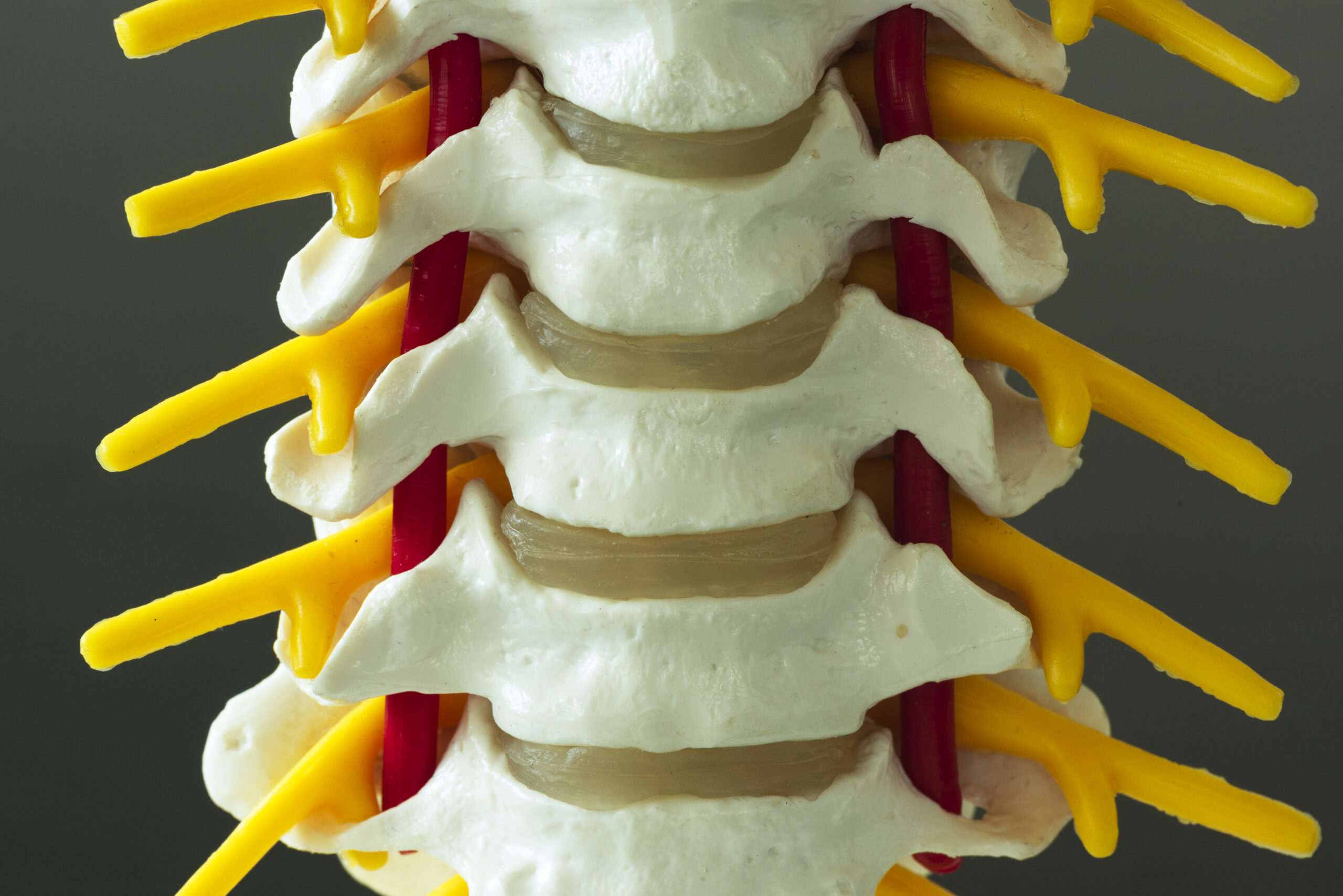
The lumbar spine anatomy consists of several interconnected components that work in harmony to provide both stability and flexibility. These components include vertebral bodies, intervertebral discs, facet joints, ligaments, muscles, and the spinal cord and nerve roots that pass through the region.
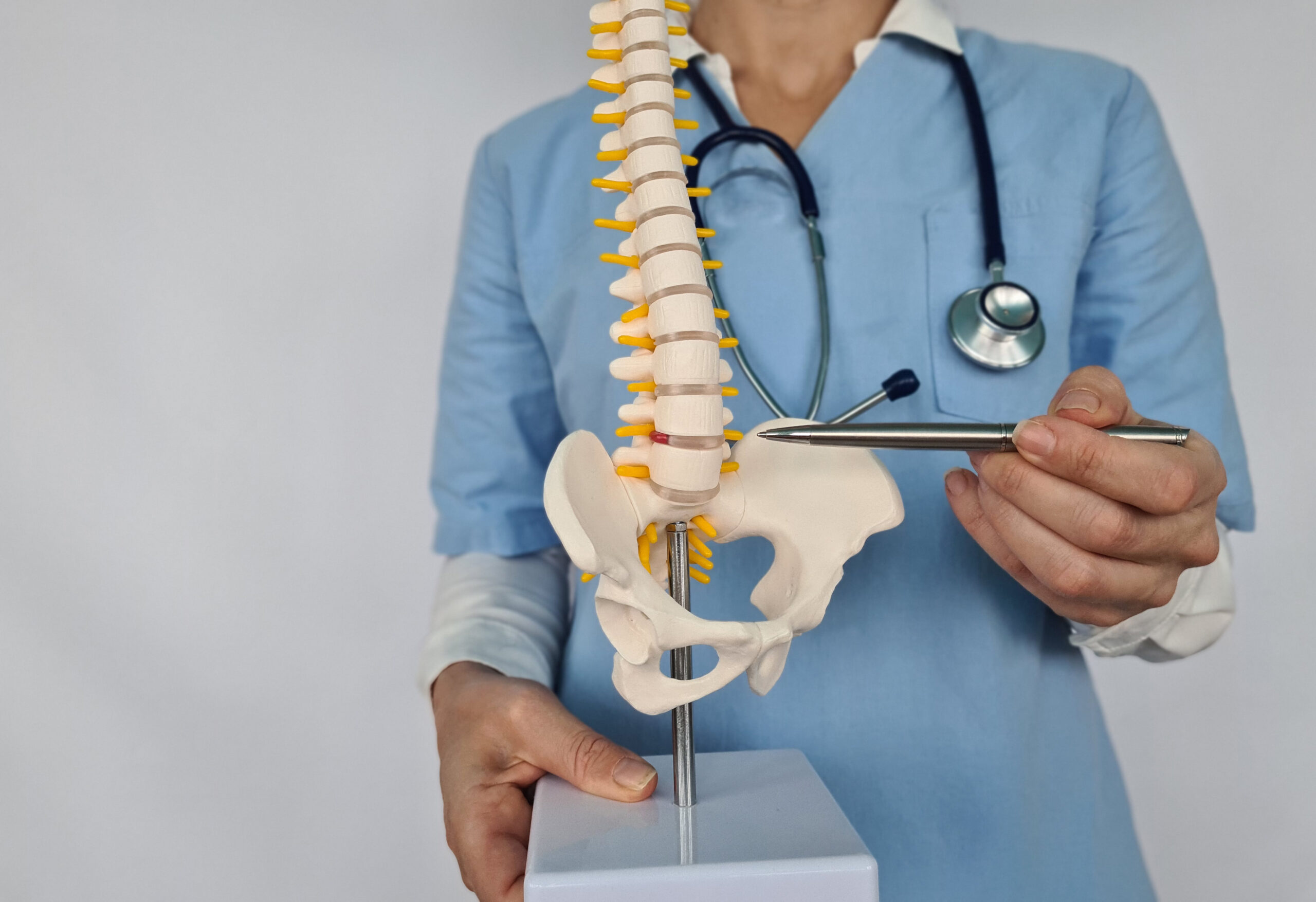
The Lower Back Vertebrae: L1 Through L5
The most defining structural feature of the lumbar spine is its five vertebrae, labeled L1 through L5. These lower back vertebrae are the largest and most robust in the entire spinal column, a design that reflects their role in supporting the upper body’s weight.
- L1 (First Lumbar Vertebra): Positioned at the top of the lumbar region, L1 marks the transition from the thoracic spine. It supports the weight transferred from the ribcage and upper body and serves as an attachment point for several important muscle groups.
- L2 (Second Lumbar Vertebra): Situated just below L1, this vertebra continues the structural support of the spinal column and forms part of the channel through which the spinal cord and nerves travel downward.
- L3 (Third Lumbar Vertebra): Located at approximately the center of the lumbar region, L3 is often described as the most centrally positioned vertebra. It plays a significant role in rotational movements of the trunk and is a common site for degenerative changes over time.
- L4 (Fourth Lumbar Vertebra): This vertebra is particularly significant in clinical practice. The L4-L5 disc level is among the most frequently affected segments in lumbar disc herniation and spinal stenosis. L4 also aligns closely with the iliac crest, making it a useful anatomical landmark in physical examinations.
- L5 (Fifth Lumbar Vertebra): As the lowest of the lower back vertebrae, L5 bears more weight than any other vertebra in the spine. It connects to the sacrum at the lumbosacral junction, a region subject to enormous mechanical forces. The L5-S1 junction is one of the most commonly affected areas in lower back disorders.
Intervertebral Discs: The Shock Absorbers
Between each pair of lumbar vertebrae lies an intervertebral disc — a fibrocartilaginous structure that acts as a cushion and shock absorber. Each disc consists of two main components: the nucleus pulposus, a soft, gel-like core that distributes pressure evenly, and the annulus fibrosus, a tough outer ring of fibrous cartilage that contains the nucleus and provides structural integrity.
These discs allow the lumbar spine to bend, flex, and rotate while protecting the vertebrae from direct impact. Unfortunately, they are also highly susceptible to degeneration with age. As the discs lose hydration and elasticity over time, conditions such as disc herniation, bulging discs, and degenerative disc disease can develop — often leading to pain that radiates into the legs, a condition commonly known as sciatica.
Facet Joints and Their Role in Spinal Movement
Each lumbar vertebra connects to its neighbors through paired facet joints, also referred to as zygapophyseal joints. These small joints are located at the back of the vertebrae and guide the direction of movement in the lumbar spine. They allow for forward and backward bending (flexion and extension) while limiting excessive rotation that might otherwise damage the spinal structures.
Facet joints are lined with cartilage and enclosed within a joint capsule filled with synovial fluid, which provides lubrication. Like other joints in the body, they can become arthritic over time, leading to a condition known as facet joint syndrome — a significant contributor to chronic lower back pain in older adults.
Ligaments and Muscles: Stability and Support
The lumbar spine is stabilized by a network of strong ligaments that connect the vertebrae and limit excessive movement. The most important of these include:
- Anterior Longitudinal Ligament (ALL): Runs along the front of the vertebral bodies, preventing hyperextension.
- Posterior Longitudinal Ligament (PLL): Runs along the back of the vertebral bodies inside the spinal canal, resisting excessive flexion and disc herniation.
- Ligamentum Flavum: Connects adjacent vertebral arches and helps maintain the curvature of the spine. Thickening of this ligament can contribute to spinal stenosis.
Additional Resources