Understanding Thoracic Outlet Syndrome and Diagnostic Challenges
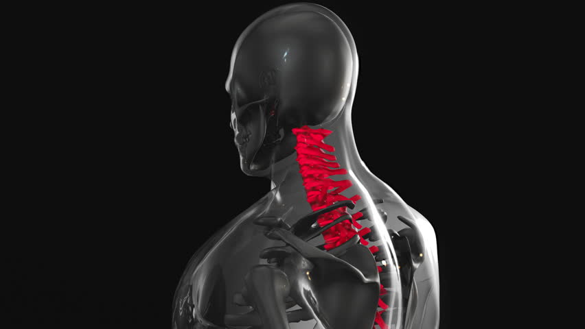
Thoracic Outlet Syndrome (TOS) occurs when nerves or blood vessels near the neck and shoulder are compressed, causing symptoms like unilateral arm numbness, pain, and weakness. Diagnosing TOS can be complex due to overlapping symptoms with conditions like cervical radiculopathy or carpal tunnel syndrome. Accurate identification requires a combination of clinical evaluation and advanced diagnostic tools. Patients often present with non-specific complaints, making it critical to differentiate TOS from other neurological or vascular issues. Early and precise diagnosis is essential to guide appropriate management, whether conservative or interventional. This article explores the most reliable imaging and orthopedic tests used to confirm TOS, emphasizing their roles in clinical decision-making.
Advanced Imaging Techniques for TOS Evaluation
Advanced imaging plays a key role in visualizing structural abnormalities associated with TOS. Magnetic Resonance Imaging (MRI) is widely used to assess soft tissues, including nerves and blood vessels, and can detect compression in the thoracic outlet. X-rays may reveal anatomical variations like cervical ribs or abnormal bone structures contributing to TOS. Computed Tomography (CT) scans, particularly with contrast, help evaluate vascular involvement, such as subclavian artery or vein compression. While these tools provide valuable insights, they are often interpreted alongside physical exams and patient history. Imaging is most effective when combined with clinical findings to confirm TOS and rule out alternative diagnoses.
Orthopedic Physical Exams and Provocation Tests
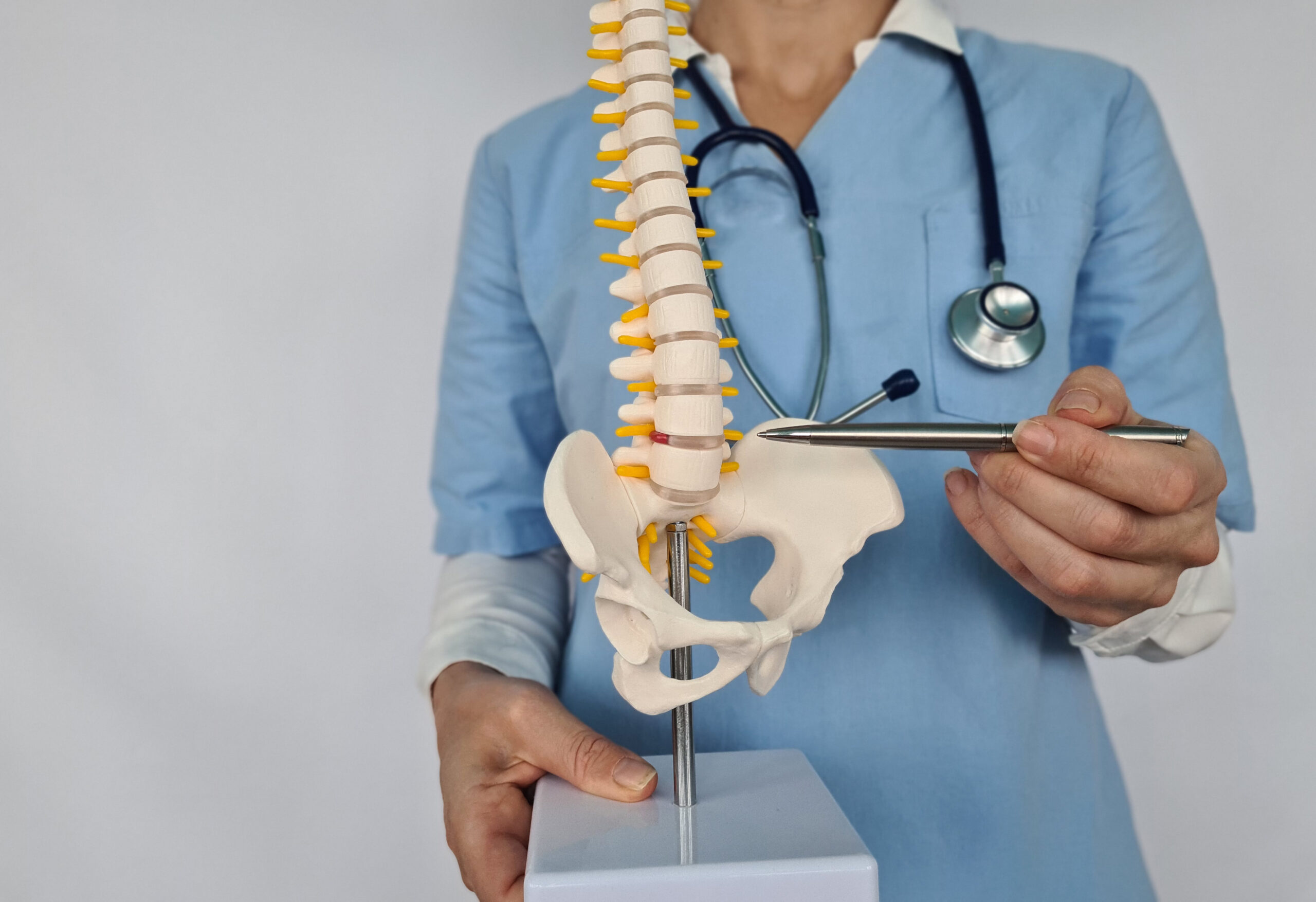
Orthopedic physical exams are foundational in TOS diagnosis. Tests like Adson’s maneuver, which assesses radial pulse changes during neck movement, and the Roos test, which evaluates symptoms during arm elevation, are commonly used. The Wright’s test and Halstead’s maneuver also help identify nerve or vascular compression. These provocative tests are non-invasive and can guide further imaging. However, their reliability varies, as results depend on examiner skill and patient cooperation. While not definitive on their own, they provide critical clues about the syndrome’s type—neurogenic, vascular, or mixed. Integrating these findings with imaging ensures a more accurate diagnosis.
Nerve Conduction Studies and Electromyography
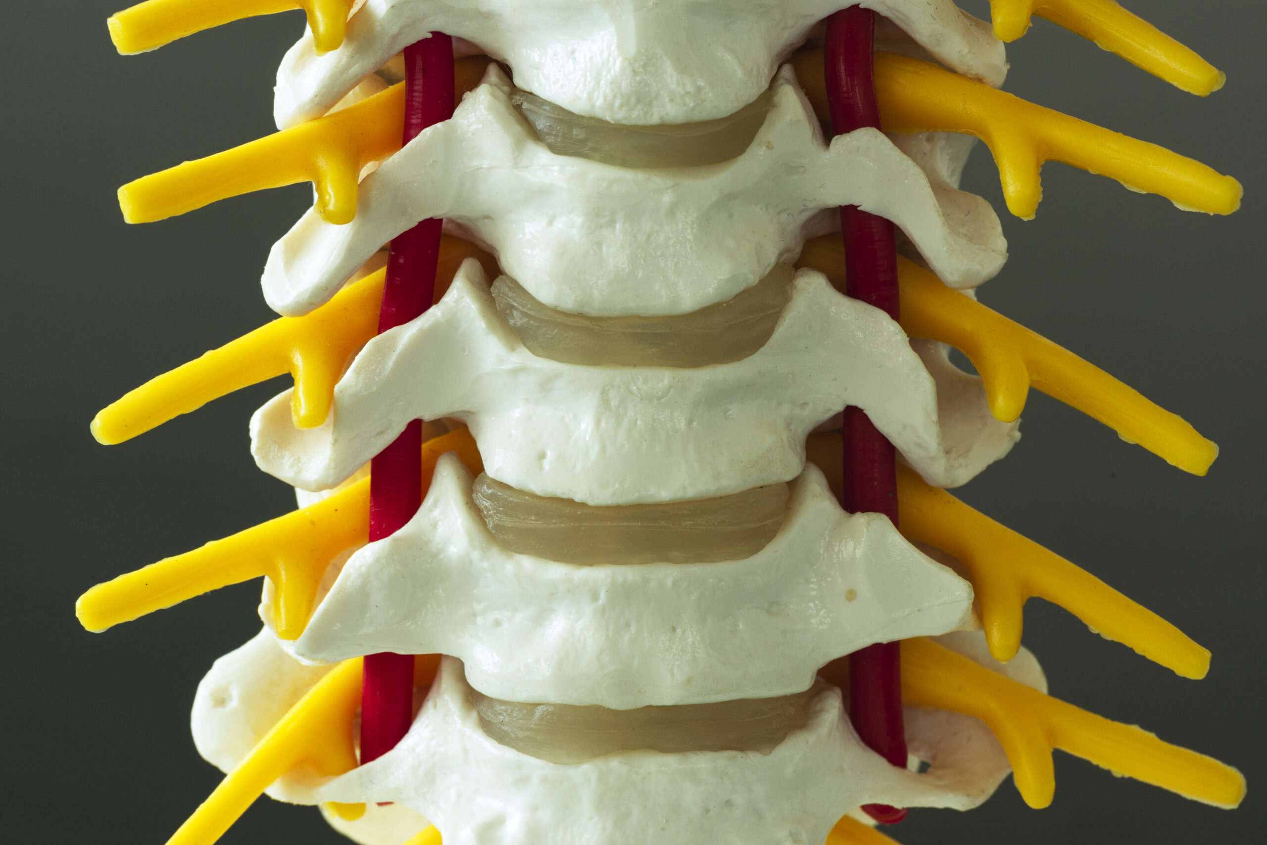
Nerve conduction studies (NCS) and electromyography (EMG) assess nerve function and muscle activity, aiding in TOS diagnosis. NCS measures how quickly nerves transmit signals, while EMG evaluates muscle response to nerve stimulation. These tests can detect abnormalities in the brachial plexus or lower cervical nerves, common in neurogenic TOS. However, they are less effective for vascular TOS, which requires imaging. NCS/EMG results must be interpreted cautiously, as they can be influenced by other conditions like peripheral neuropathy. When combined with physical exams and imaging, they enhance diagnostic accuracy, particularly in complex cases where symptoms are atypical.
Limitations and Considerations in Diagnostic Testing
Despite their utility, diagnostic tests for TOS have limitations. Imaging may miss subtle compressions, and physical exams can yield false positives or negatives. Patient factors like obesity or prior surgeries can complicate interpretations. Additionally, TOS symptoms often mimic other disorders, leading to diagnostic delays. Clinicians must balance test results with clinical judgment, avoiding over-reliance on any single method. A multidisciplinary approach, involving specialists like neurologists or vascular surgeons, may be necessary for ambiguous cases. Patient history and symptom patterns remain vital in guiding the diagnostic process, ensuring tests are used strategically.
Integrating Diagnostic Tools for Accurate TOS Identification
Diagnosing TOS requires a tailored, integrated approach. Advanced imaging, orthopedic exams, and nerve studies each contribute unique insights but are most effective when used collectively. For example, MRI might confirm soft tissue compression, while physical tests suggest nerve involvement. Clinicians must weigh test results against clinical context, avoiding overdiagnosis or missed cases. Patient-reported symptoms, such as activity-related numbness or nighttime discomfort, further inform decisions. By combining objective data with clinical expertise, healthcare providers can enhance diagnostic precision. This comprehensive strategy ensures patients receive targeted care, whether through conservative management or specialized interventions.