Anatomy of the Thoracic Outlet and Key Structures
The thoracic outlet is a narrow space between the base of the neck and the armpit, bounded by the clavicle, first rib, and scalene muscles. This region houses critical structures, including the brachial plexus (a network of nerves), subclavian artery, and subclavian vein. Understanding this anatomy is vital for chiropractors, as compression of these elements can lead to thoracic outlet syndrome (TOS). The cervical spine, particularly the seventh cervical vertebra (C7), and the first rib play central roles in maintaining the space for these structures. Misalignments or abnormalities in these areas can alter the thoracic outlet's dimensions, potentially causing symptoms like pain, numbness, or circulation issues in the arm. Chiropractors must recognize how variations in spinal curvature, rib positioning, or musculature contribute to TOS, enabling targeted interventions to restore balance and function.
Role of the Cervical Spine in TOS
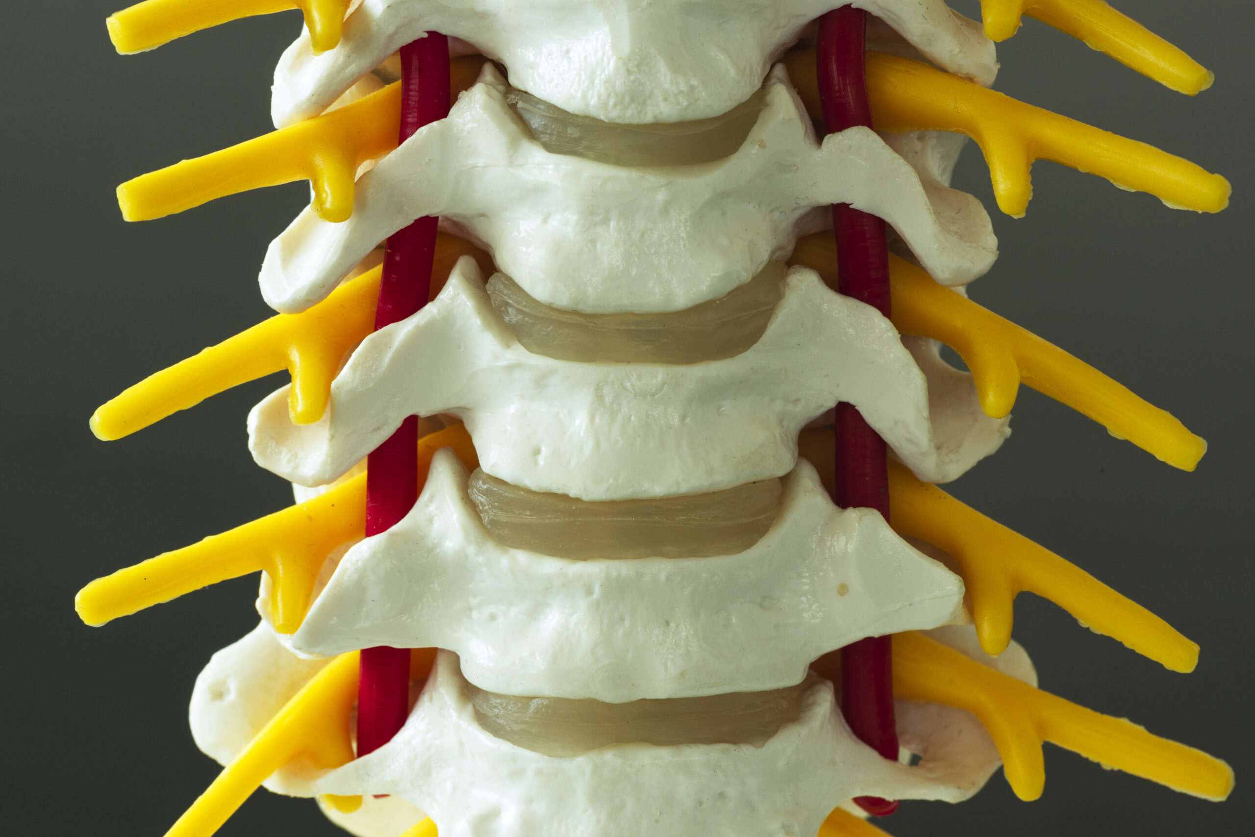
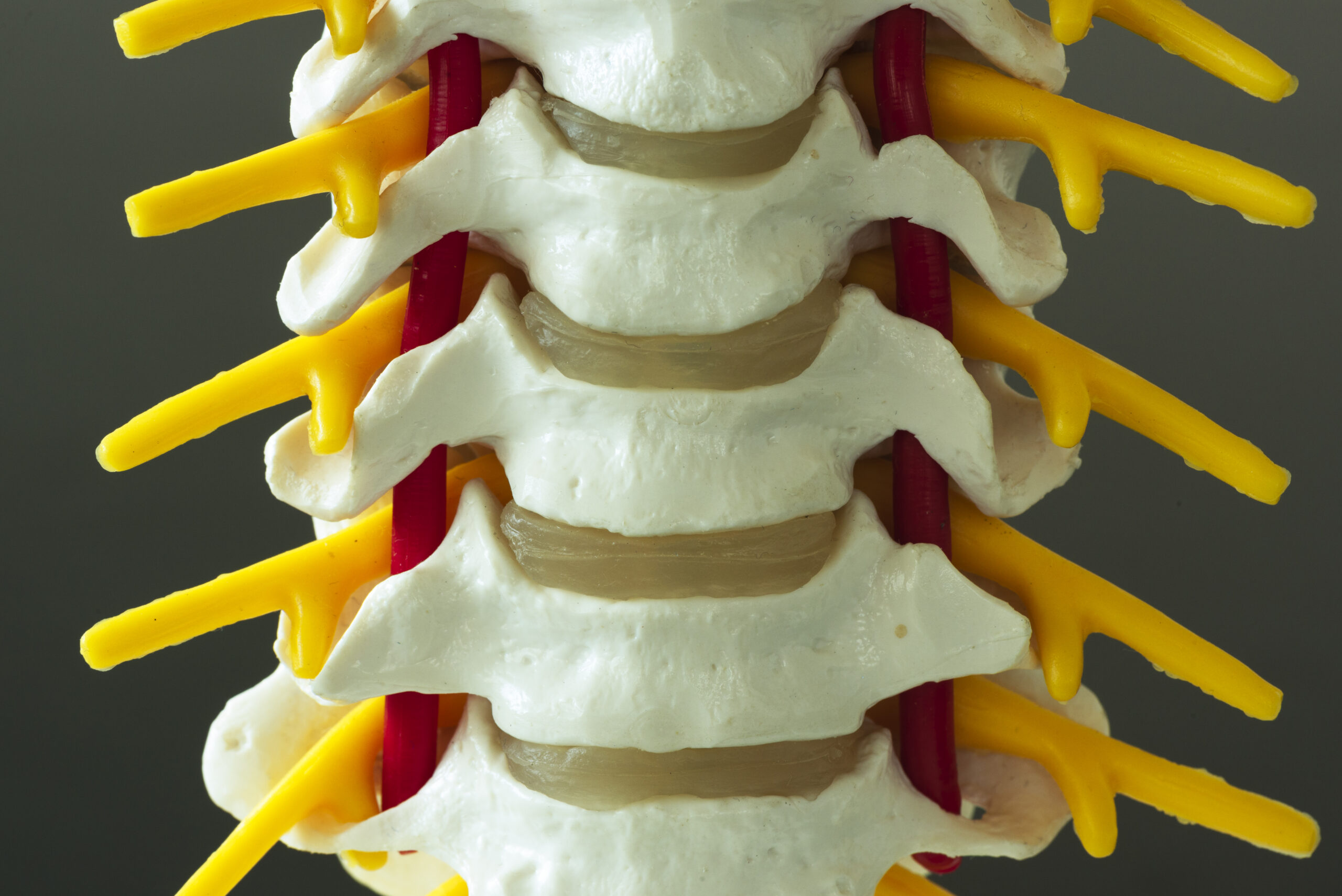
The cervical spine, especially the lower vertebrae (C5-C8), directly influences the brachial plexus and vascular pathways. Abnormalities such as herniated discs, facet joint dysfunction, or spinal stenosis can compress nerves or blood vessels, mimicking TOS symptoms. For example, a bulging disc at C7 might irritate the lower brachial plexus, causing arm pain or weakness. Chiropractors must assess cervical mobility and alignment, as restricted movement or subluxations can exacerbate thoracic outlet compression. Additionally, the cervical curve's integrity affects overall posture, which indirectly impacts the thoracic outlet. By addressing cervical spine health, chiropractors can alleviate secondary stress on the thoracic region, reducing the risk of nerve or vascular compromise. This underscores the need for a holistic approach to spinal care in TOS management.
First Rib and Scapular Mechanics
The first rib and scapula (shoulder blade) form a dynamic relationship critical to thoracic outlet function. The first rib's position and mobility are essential for maintaining adequate space for the brachial plexus and subclavian vessels. A displaced or elevated first rib can narrow this space, leading to compression. Chiropractors must evaluate the rib's alignment, as even minor deviations can disrupt nerve and vascular flow. The scapula's movement also plays a role; altered mechanics, such as winging or protraction, may alter the thoracic outlet's geometry. For instance, a forward-rounded shoulder posture can compress the subclavian artery. Understanding these interactions allows chiropractors to employ techniques like rib adjustments or scapular mobilization to restore optimal positioning. This knowledge is foundational for addressing mechanical contributors to TOS and improving patient outcomes through precise, targeted interventions.
Muscular Contributions to Compression
Several muscles surrounding the thoracic outlet can contribute to compression if tight, imbalanced, or overactive. The scalene muscles (anterior, middle, and posterior) are particularly significant, as they form part of the thoracic outlet's boundary. Tightness in these muscles can compress the brachial plexus or subclavian artery, mimicking TOS symptoms. Similarly, the pectoralis minor muscle, located in the upper chest, can pull the clavicle downward, narrowing the thoracic outlet. Chiropractors must assess muscle tone and function, as myofascial restrictions or trigger points can perpetuate compression. Techniques like soft tissue therapy, stretching, or myofascial release may be employed to alleviate tension. Additionally, weakness in stabilizing muscles, such as the serratus anterior, can lead to scapular instability, further complicating the condition. Recognizing these muscular dynamics enables chiropractors to develop comprehensive treatment plans that address both structural and functional components of TOS.
Neurological and Vascular Pathways Involved
The brachial plexus and vascular structures in the thoracic outlet are complex networks with distinct pathways. The brachial plexus originates from the cervical spinal nerves (C5-T1) and travels through the thoracic outlet to innervate the arm. Compression at any point along this pathway can cause symptoms like tingling, numbness, or weakness. Similarly, the subclavian artery and vein pass through the thoracic outlet, and their compression may lead to reduced blood flow, causing arm fatigue or discoloration. Chiropractors must understand these pathways to differentiate TOS from other conditions with overlapping symptoms. For example, a cervical radiculopathy might present similarly but involves nerve root compression rather than thoracic outlet issues. By mapping these anatomical routes, chiropractors can identify the precise location of dysfunction and tailor adjustments or therapies to relieve pressure on nerves and vessels effectively.
Clinical Implications for Chiropractic Adjustments
Chiropractors treating TOS must integrate anatomical knowledge into their clinical practice. Adjustments to the cervical spine, first rib, or thoracic vertebrae can alleviate compression by restoring proper alignment and mobility. For instance, a cervical spine adjustment may reduce nerve irritation, while a first rib manipulation can widen the thoracic outlet. Soft tissue techniques targeting tight muscles like the scalenes or pectoralis minor complement these adjustments by addressing muscular contributors to compression. Additionally, posture correction and rehabilitative exercises are essential for long-term management, as they prevent recurrence. Chiropractors must also consider the patient's overall biomechanics, such as shoulder girdle alignment or spinal curvature, when developing treatment plans. By combining structural and functional approaches, chiropractors can effectively manage TOS, emphasizing the importance of a thorough understanding of spinal anatomy in clinical decision-making.