The Role of Thoracic Vertebrae Structure in Postural Stability
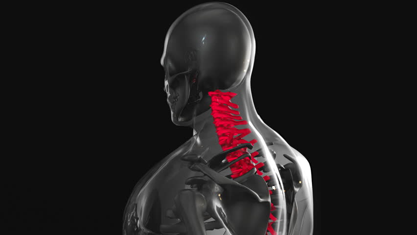
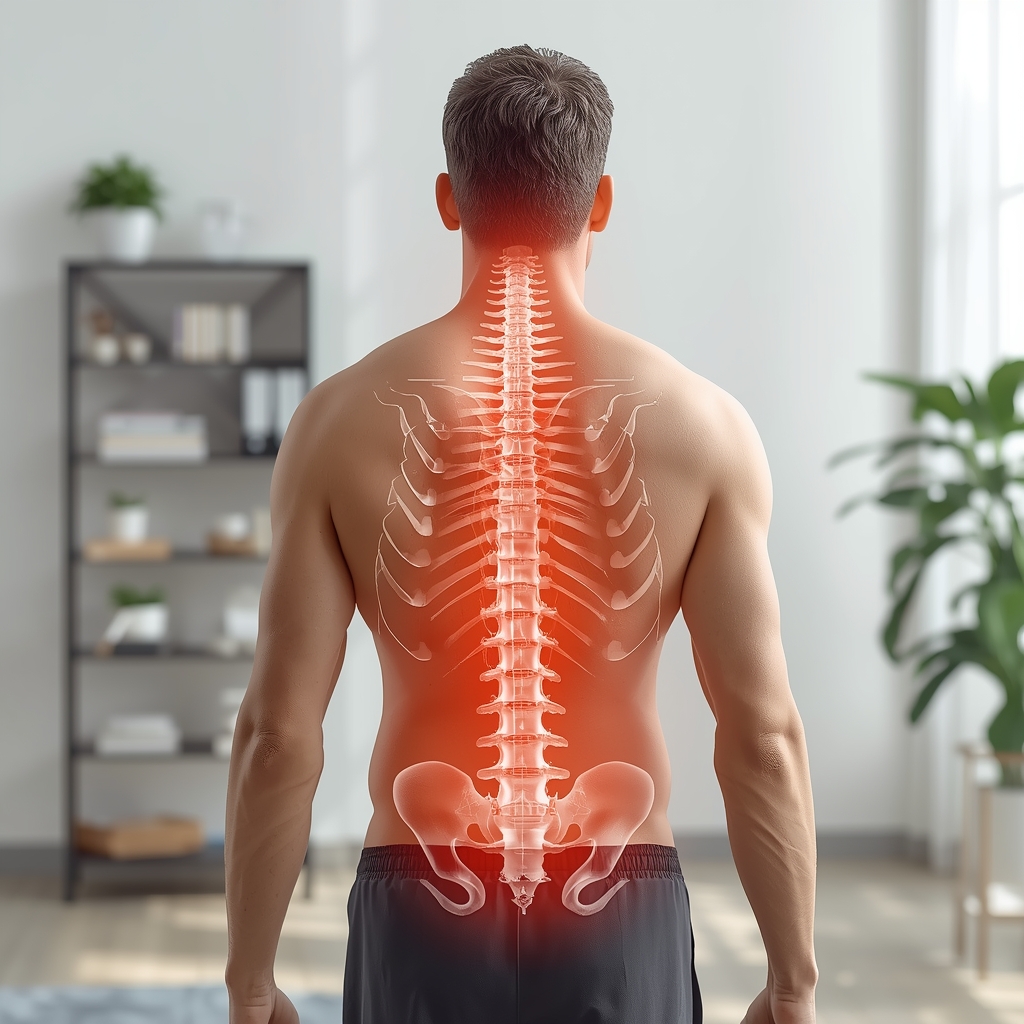
The thoracic spine consists of 12 vertebrae (T1-T12) that form the central axis of the upper body. Each vertebra has unique features, including heart-shaped vertebral bodies, short pedicles, and long, downward-pointing spinous processes. These characteristics create a natural kyphotic curve that stabilizes the rib cage and protects vital organs. The thoracic vertebrae also feature costal facets for rib articulation, which directly influence thoracic mobility and breathing mechanics. Chiropractors must understand how variations in vertebral alignment—such as excessive kyphosis or hypokyphosis—can disrupt postural balance. For example, a flattened thoracic curve may lead to forward head posture, while increased kyphosis can cause rib cage compression. Recognizing these structural relationships allows practitioners to identify misalignments that contribute to chronic postural strain, guiding adjustments and rehabilitative strategies.
Intervertebral Discs and Their Impact on Thoracic Mobility
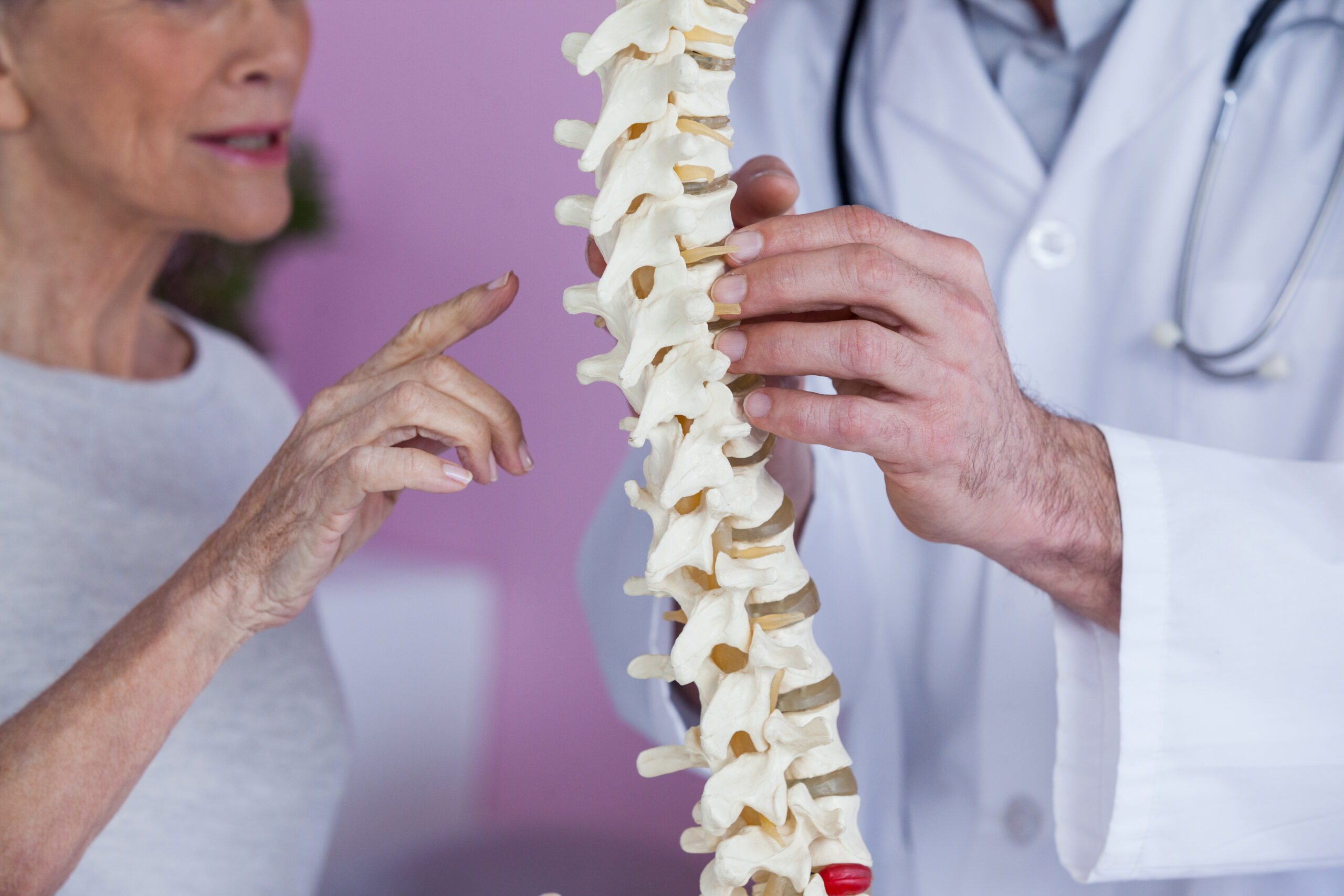
The intervertebral discs between thoracic vertebrae act as shock absorbers and facilitate flexibility. Unlike the lumbar spine, the thoracic discs have a thinner annulus fibrosus and a more gelatinous nucleus pulposus, limiting rotational movement but enhancing stability. Degenerative changes in these discs—such as dehydration or herniation—can alter spinal mechanics, leading to restricted motion and compensatory postural adaptations. Chiropractors assess disc health through range-of-motion tests and palpation, as disc-related dysfunction often manifests as localized tenderness or reduced thoracic extension. Addressing disc imbalances requires precise adjustments to restore normal curvature and prevent secondary issues like rib dysfunction or nerve irritation. Understanding disc anatomy ensures chiropractors differentiate between mechanical and pathological causes of postural deviations, tailoring interventions to each patient’s needs.
Facet Joints and Their Contribution to Thoracic Alignment
The thoracic spine’s facet joints, located between the superior and inferior articular processes of adjacent vertebrae, govern rotational and gliding movements. These synovial joints are oriented in a way that restricts excessive motion, emphasizing the thoracic spine’s role as a stabilizing segment. When facet joints become hypomobile due to injury, arthritis, or muscle tightness, they can cause compensatory shifts in posture, such as lateral curvature or uneven weight distribution. Chiropractors evaluate facet joint function through specific maneuvers, like the thoracic rotation test, to identify restrictions. Adjusting these joints requires careful technique to avoid overcorrection, as the thoracic spine’s limited mobility makes it susceptible to iatrogenic injury. By restoring facet joint mobility, practitioners help patients achieve balanced postures and reduce strain on surrounding musculature and neural structures.
The Significance of Costovertebral Joints in Postural Mechanics
The costovertebral joints, where the thoracic vertebrae connect to the ribs, play a critical role in thoracic stability and respiratory function. These joints allow for slight movement during breathing, and dysfunction here can lead to restricted thoracic expansion and altered posture. For instance, rib misalignment may cause the thoracic spine to compensate by increasing its kyphotic curve, leading to a hunched appearance. Chiropractors often address costovertebral joint dysfunctions through targeted adjustments and soft tissue work, ensuring proper rib positioning. This is particularly important in patients with postural imbalances caused by prolonged sitting or poor ergonomics. By integrating joint mobility assessments with postural analysis, practitioners can correct rib-thoracic spine relationships, improving both structural alignment and functional breathing patterns.
Thoracic Curve Variations and Their Diagnostic Implications
The thoracic spine’s natural kyphotic curve typically ranges from 20-40 degrees, providing structural support and protecting the spinal cord. Abnormalities in this curve—such as hyperkyphosis (excessive rounding) or hypokyphosis (flattened curve)—can indicate underlying postural imbalances or systemic conditions. Hyperkyphosis is often linked to poor posture, osteoporosis, or muscle weakness, while hypokyphosis may result from compensatory changes after lumbar spine interventions. Chiropractors use visual inspection, palpation, and imaging (when necessary) to assess curve integrity. Correcting these deviations involves a combination of spinal adjustments, postural re-education, and targeted exercises. Understanding the biomechanics of the thoracic curve enables practitioners to design comprehensive treatment plans that address both structural and functional aspects of postural health.
Musculature Surrounding the Thoracic Spine and Postural Control
The thoracic spine is stabilized by a complex network of muscles, including the rhomboids, trapezius, erector spinae, and serratus posterior. These muscles work synergistically to maintain posture, support the rib cage, and facilitate movement. Weakness or tightness in these muscles can lead to postural deviations, such as rounded shoulders or a forward head position. Chiropractors evaluate muscle tone and strength through manual resistance tests and palpation, identifying areas of imbalance. Treatment often includes soft tissue therapy, corrective exercises, and adjustments to restore neuromuscular coordination. By addressing both structural and muscular components, practitioners help patients achieve long-term postural stability. This holistic approach ensures that interventions target the root causes of imbalances rather than just surface symptoms.