Cervical Spine Structure and Nerve Root Anatomy
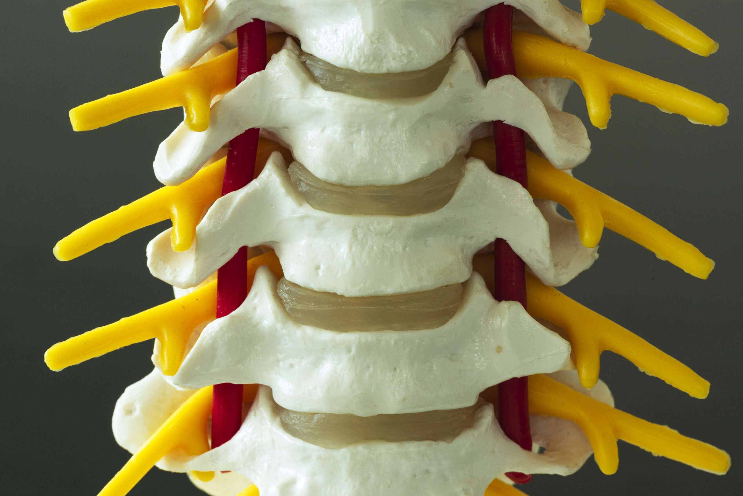
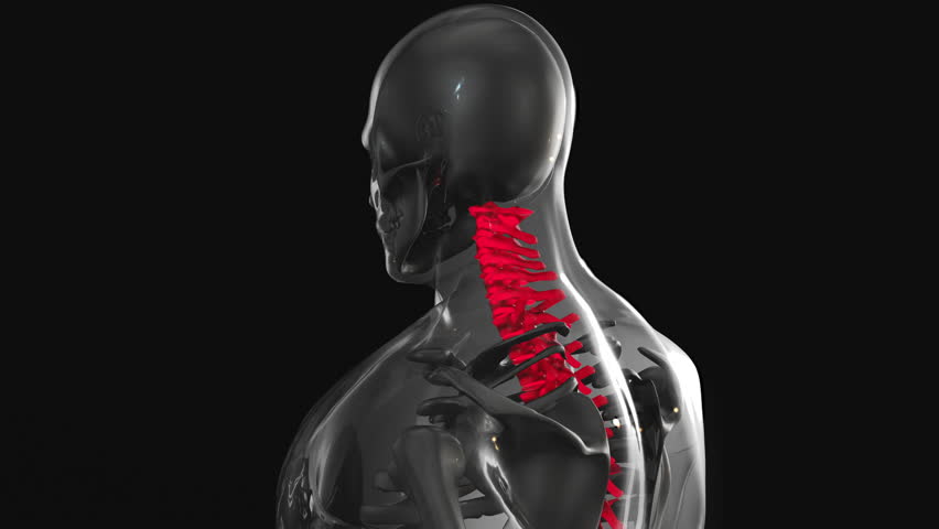
The cervical spine consists of seven vertebrae (C1-C7) that form the neck region, supporting head movement while protecting the spinal cord. Each vertebra has a central canal for the spinal cord and intervertebral foramina—openings between adjacent vertebrae—through which spinal nerves exit. These nerves, including the C5-C8 and T1 roots, branch into peripheral nerves that innervate the upper extremities. Understanding this anatomy is critical for chiropractors, as cervical radiculopathy often arises from compression or irritation of these nerves due to herniated discs, bone spurs, or degenerative changes. Recognizing the precise location of nerve exits helps identify which root is affected, guiding diagnostic and therapeutic decisions. For example, C6 radiculopathy typically causes thumb and index finger symptoms, while C8 involvement may affect the little finger. This foundational knowledge ensures accurate assessment of nerve pathways and potential sources of dysfunction.
Key Anatomical Features Influencing Cervical Radiculopathy
Several anatomical factors contribute to cervical radiculopathy. The intervertebral discs, which act as shock absorbers, can herniate and compress adjacent nerves. Facet joints, which stabilize the spine, may develop osteophytes (bone spurs) that narrow the foramina, leading to nerve entrapment. The spinal cord itself is vulnerable to compression in cases of spinal stenosis, though radiculopathy more commonly involves nerve roots. Additionally, the brachial plexus—formed by C5-T1 nerves—transmits signals to the arms, making it a target for radiculopathy. Chiropractors must differentiate between radiculopathy and other conditions, such as thoracic outlet syndrome, which affects the same nerve pathways. By mapping these structures, chiropractors can pinpoint the origin of symptoms, whether it’s a disc issue, joint dysfunction, or soft tissue involvement. This precision is essential for tailoring interventions that address the root cause rather than just alleviating symptoms.
Clinical Implications for Chiropractic Diagnosis
Chiropractors rely on anatomical knowledge to interpret patient symptoms and perform targeted exams. For instance, a patient reporting arm pain, numbness, or weakness may have radiculopathy, but the specific nerve root involved determines the diagnostic approach. Physical tests, such as the Spurling’s test (which reproduces symptoms by extending and rotating the neck), help confirm nerve compression. Imaging studies like X-rays or MRI may also be used to visualize disc herniations or spinal stenosis. However, chiropractors often prioritize clinical correlation over imaging, as anatomical understanding allows them to assess posture, range of motion, and muscle strength. For example, a weakened biceps reflex might indicate C5-C6 involvement, while diminished triceps reflex suggests C7-C8 compression. This anatomically informed diagnosis ensures that treatment plans address the specific structures contributing to the patient’s condition.
Treatment Approaches Based on Anatomical Principles
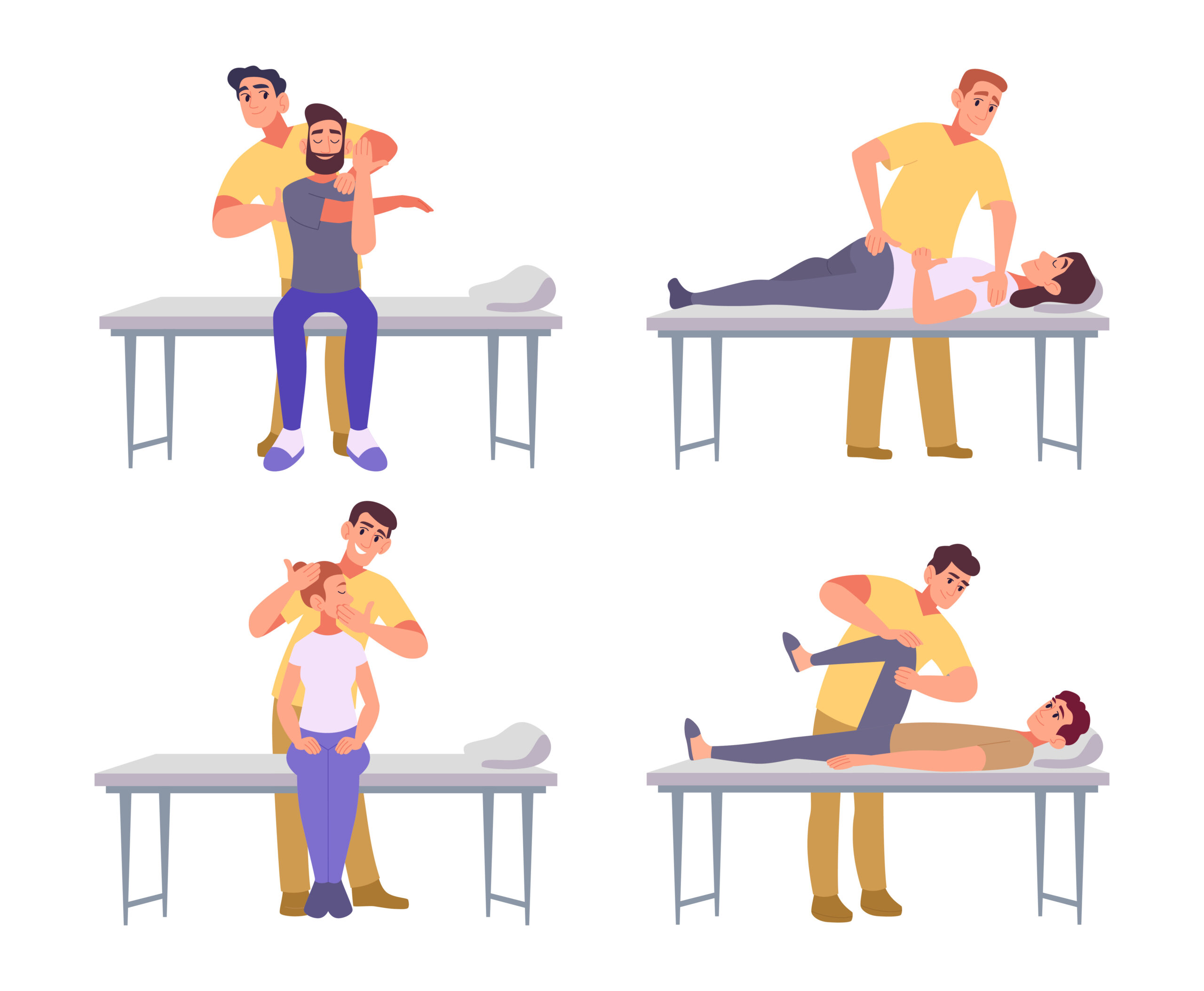
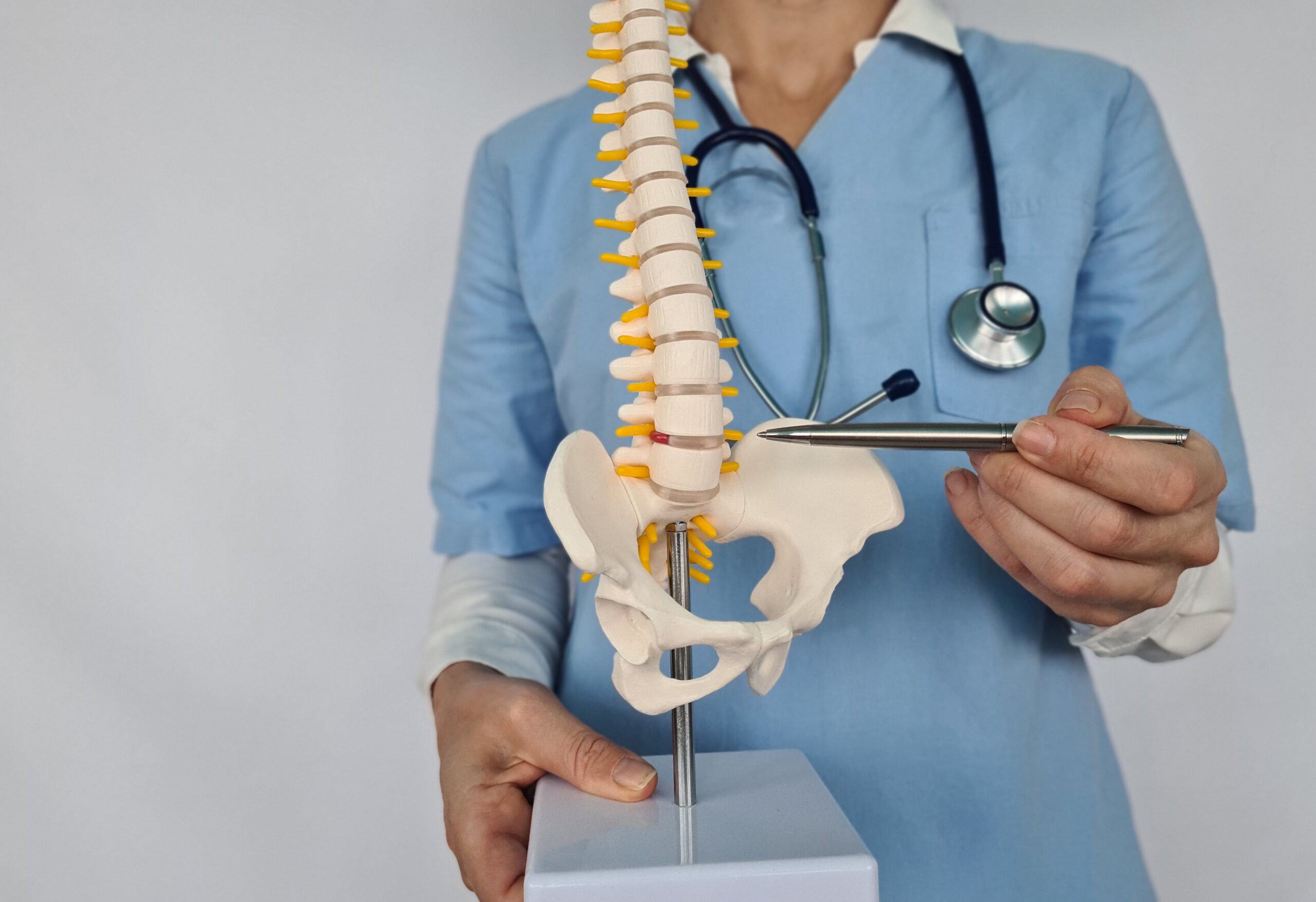
Chiropractic care for cervical radiculopathy focuses on reducing nerve compression and restoring normal spinal function. Spinal adjustments aim to realign vertebrae, alleviating pressure on affected nerves. For example, a misaligned C6 vertebra may be corrected to relieve symptoms in the thumb and index finger. Decompression techniques, such as flexion-distraction, gently stretch the spine to create space for herniated discs, reducing nerve irritation. Soft tissue therapies, like myofascial release, address muscle tension that exacerbates nerve compression. Chiropractors also consider postural habits, recommending ergonomic adjustments or exercises to prevent recurrence. However, treatment plans are individualized, as each patient’s anatomy and condition vary. Collaboration with other healthcare providers may be necessary for complex cases. The goal is to harmonize anatomical understanding with clinical judgment to achieve optimal outcomes without invasive interventions.
Soft Tissue and Postural Contributions to Cervical Radiculopathy
While bony structures are central to cervical radiculopathy, soft tissues and posture play significant roles. Tightness in the levator scapulae or suboccipital muscles can alter cervical alignment, increasing nerve root vulnerability. Poor posture, such as forward head positioning, narrows the intervertebral foramina, heightening the risk of compression. Chiropractors assess these factors during evaluations, as they may contribute to or perpetuate radiculopathy. For example, a patient with chronic neck strain might benefit from stretching exercises and postural retraining alongside spinal adjustments. Soft tissue techniques, such as trigger point therapy, can relieve muscle tension that exacerbates nerve irritation. Addressing these biomechanical imbalances is crucial for long-term relief, as isolated spinal adjustments may not resolve underlying issues. Integrating soft tissue and postural strategies ensures a comprehensive approach that aligns with the patient’s anatomical needs.
Integrating Anatomy with Patient Education and Prevention
Chiropractors use anatomical knowledge to educate patients about their condition and empower self-management. Explaining how nerve compression occurs—such as a herniated disc pressing on a root—helps patients understand their symptoms and treatment rationale. This education also emphasizes preventive measures, like maintaining proper posture and avoiding repetitive neck strain. For instance, patients may learn to adjust their workspace to reduce forward head posture, which strains cervical structures. Chiropractors might also recommend exercises to strengthen neck muscles and improve flexibility, reducing future risks. By linking anatomical concepts to daily habits, patients gain insights into how their body functions and how to protect it. This proactive approach not only supports recovery but also fosters long-term musculoskeletal health, aligning care with the patient’s lifestyle and goals.