Understanding Thermography in Chiropractic Care
Chiropractic thermography is a non-invasive diagnostic tool that has steadily gained recognition within the chiropractic profession. By measuring temperature variations across the skin’s surface, this technology allows practitioners to identify areas of physiological stress, nerve interference, and inflammation that may not be visible through conventional examination methods. For patients seeking a more comprehensive understanding of their spinal health, thermography offers a window into the body’s neurological and musculoskeletal function that is both detailed and revealing.
At its core, chiropractic thermography relies on the principle that the nervous system regulates blood flow and skin temperature throughout the body. When nerve function is compromised — whether due to spinal misalignment, soft tissue injury, or chronic inflammation — temperature asymmetries emerge on the surface of the skin. Detecting these patterns with precision is where infrared imaging chiropractic technology excels.
How Does Infrared Imaging Work in a Chiropractic Setting?
Infrared imaging in chiropractic practice involves the use of highly sensitive thermal cameras that capture heat emitted naturally from the body. Unlike X-rays, which examine bone structure, or MRI scans, which assess soft tissue in detail, infrared thermography evaluates the functional state of the nervous system by measuring radiated heat. The procedure is completely passive — no radiation is emitted, no contact is required, and no preparation beyond temperature acclimatisation is necessary.
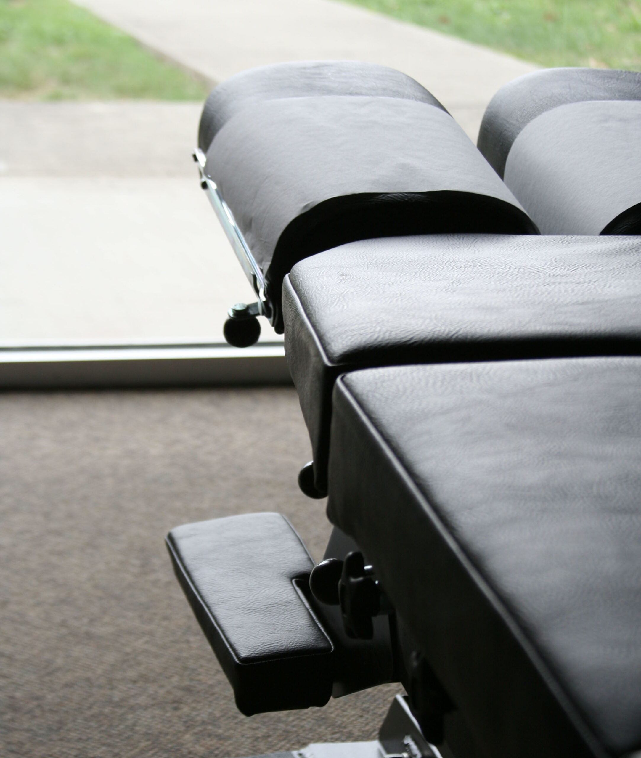
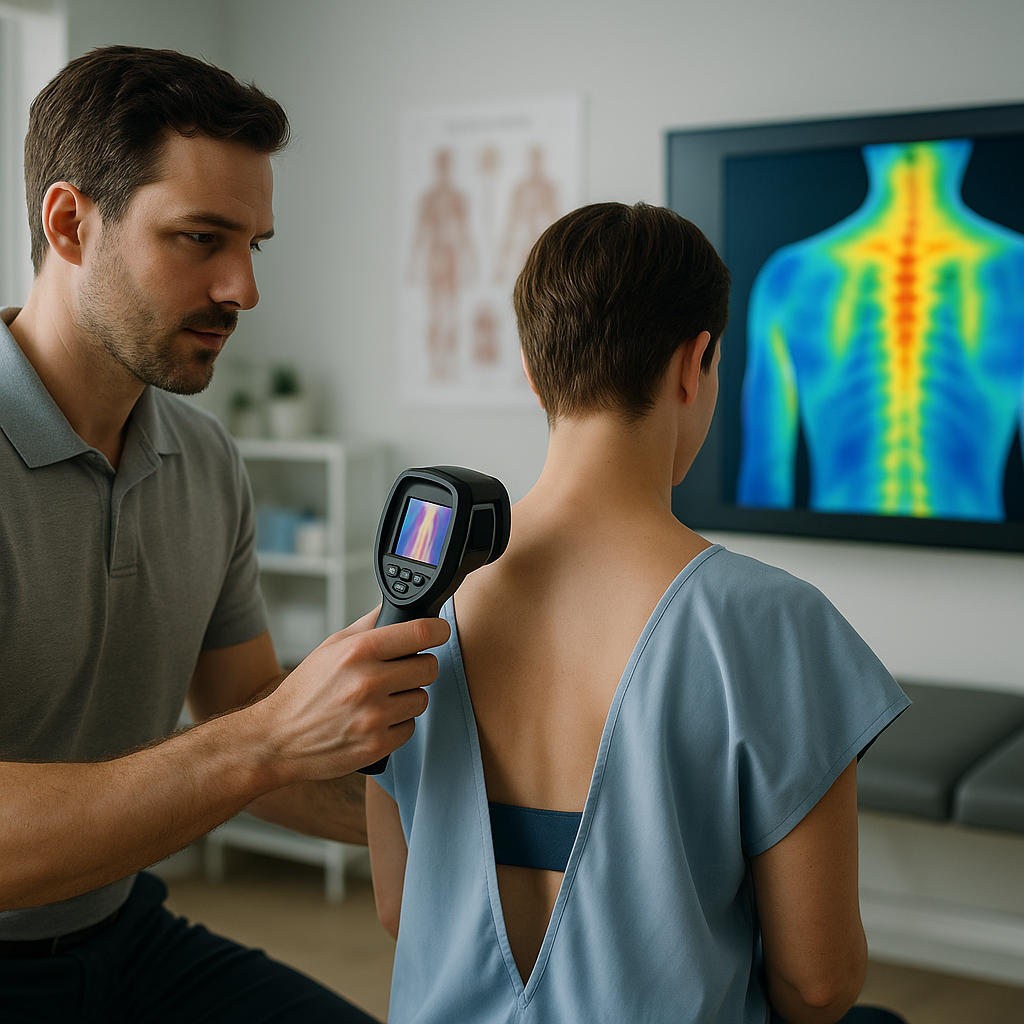
During a session, the patient stands or sits in a temperature-controlled room for a brief period to allow the skin to reach thermal equilibrium with the environment. The practitioner then uses a calibrated infrared camera to capture thermal images of targeted regions — most commonly the spine and surrounding musculature. The resulting images are displayed as colour-coded maps, where cooler areas typically appear in blue or green tones and warmer areas appear in orange or red. These colour gradients allow the chiropractor to identify patterns of thermal asymmetry that may indicate underlying dysfunction.
What Is a Thermal Spine Scan?
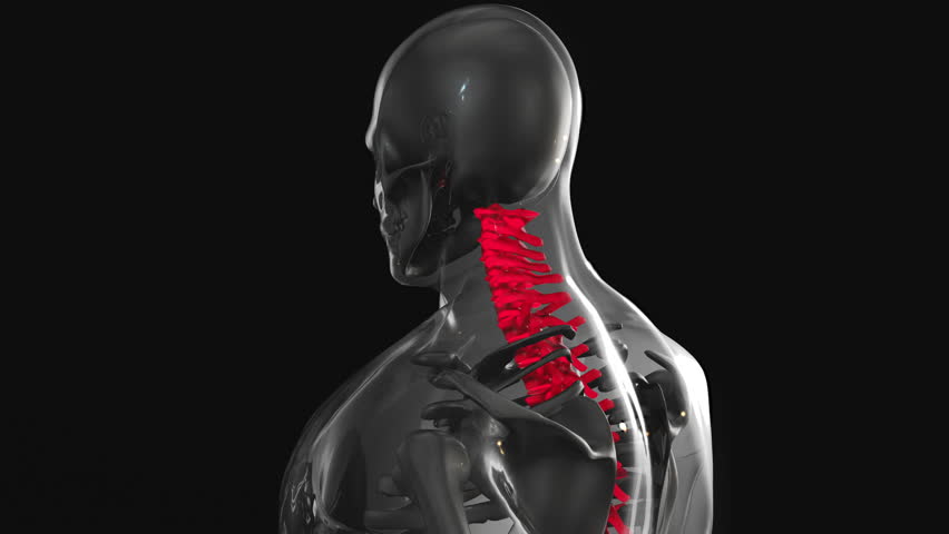
A thermal spine scan is one of the most common applications of thermography within chiropractic practice. It involves capturing sequential thermal images along the length of the spine to detect temperature differences between the left and right sides of the paraspinal region — the area immediately flanking the vertebral column.
Healthy spinal function tends to produce symmetrical temperature patterns on both sides of the spine. When subluxations — areas of vertebral misalignment that interfere with nerve communication — are present, they often produce characteristic temperature differentials. These deviations serve as objective indicators that help chiropractors locate areas requiring adjustment and assess the severity of nerve involvement.
The thermal spine scan is particularly valued because it provides a functional picture of how the nervous system is performing along the entire spinal column. This makes it a useful complement to structural assessments and helps practitioners tailor their treatment strategies with greater precision. It is also frequently used as a monitoring tool, allowing both the chiropractor and the patient to track changes in nerve function over the course of care.
The Role of Heat Pattern Analysis in Diagnosis
Heat pattern analysis is the interpretive component of thermographic examination. Once thermal images have been captured, the chiropractor analyses the distribution and intensity of heat patterns to draw clinical conclusions. This process requires both technological precision and a thorough understanding of neuroanatomy, as the patterns observed must be correlated with specific spinal levels and their associated nerve pathways.
Several key indicators are examined during heat pattern analysis:
- Thermal asymmetry: Differences in temperature between corresponding areas on opposite sides of the body may indicate nerve irritation, vascular changes, or inflammatory activity.
- Hot spots: Localised areas of elevated temperature often point to acute inflammation, increased metabolic activity, or nerve irritation in that region.
- Cold zones: Areas of reduced temperature may suggest compromised blood flow, nerve inhibition, or chronic degeneration.
- Pattern consistency: Recurring thermal patterns across multiple scans can indicate chronic conditions or persistent subluxation complexes that require ongoing attention.
By evaluating these variables in conjunction with patient history and physical examination findings, chiropractors can develop a more complete clinical picture. This multi-faceted approach enhances the accuracy of diagnosis and supports more targeted and effective treatment planning.
Primary Uses of Thermography in Chiropractic Practice
Chiropractic thermography serves a wide range of clinical purposes. Its applications extend beyond initial diagnosis and have become integral to patient management in many progressive chiropractic clinics. The following represent the most significant ways in which thermography is applied in practice:
1. Detecting Vertebral Subluxations
One of the foundational applications of chiropractic thermography is identifying vertebral subluxations — misalignments of the spinal vertebrae that interfere with the nervous system’s ability to communicate efficiently throughout the body. Because subluxations affect nerve function, they frequently produce measurable changes in skin temperature. Thermal scanning enables chiropractors to pinpoint these areas with a level of objectivity that complements other diagnostic tools.
2. Assessing Neurological Function
The nervous system’s influence over circulation and thermal regulation means that infrared imaging provides a reliable indirect measure of neurological activity. Spinal nerve roots control vasomotor tone in the skin, and disruptions to these pathways result in detectable temperature changes. Thermography, therefore, offers a non-invasive means of assessing how well the nervous system is functioning at different levels of the spine.
3. Monitoring Treatment Progress
Perhaps one of the most clinically valuable uses of thermographic scanning is its role in evaluating the effectiveness of chiropractic adjustments over time. By comparing thermal images taken before, during, and after a course of treatment, practitioners can objectively demonstrate changes in nerve function and inflammation levels. This evidence-based approach not only guides clinical decision-making but also provides patients with tangible data regarding their progress.