Understanding MRI in the Context of Chiropractic Care
Magnetic resonance imaging (MRI) has become one of the most powerful diagnostic tools available in modern healthcare. While many people associate MRI scans with hospital-based specialties such as neurology or orthopedic surgery, the role of chiropractic MRI is increasingly significant. Chiropractors are trained musculoskeletal specialists, and advanced imaging plays a critical part in helping them deliver precise, evidence-based care to their patients.
This article explores how magnetic resonance imaging chiropractic applications work, why MRI is sometimes necessary in a chiropractic setting, and what patients can expect when their chiropractor recommends this form of imaging.
What Is MRI and How Does It Work?
Magnetic resonance imaging is a non-invasive diagnostic technique that uses powerful magnetic fields and radio waves to produce detailed images of the body’s internal structures. Unlike X-rays or CT scans, MRI does not use ionizing radiation, making it a safer option for visualizing soft tissues such as muscles, ligaments, intervertebral discs, and spinal cord structures.
The images produced by MRI are exceptionally detailed, allowing healthcare providers to observe subtle changes in tissue that would otherwise be invisible through conventional radiography. This level of detail is especially valuable when evaluating complex spinal conditions, which are among the most common reasons patients seek chiropractic care.
Why Chiropractors May Recommend an MRI
Chiropractors are trained to assess, diagnose, and treat conditions of the musculoskeletal and nervous systems. In many cases, a thorough physical examination and standard X-rays are sufficient to guide treatment. However, there are specific clinical scenarios where MRI spine diagnosis becomes essential. These include:
- Suspected disc herniation or disc degeneration: MRI provides unparalleled visualization of intervertebral discs, helping to confirm whether a herniated disc is compressing nearby nerve roots.
- Radiculopathy or nerve compression: When a patient presents with radiating pain, numbness, or tingling down the arms or legs, MRI can identify the precise location and cause of nerve involvement.
- Spinal stenosis: Narrowing of the spinal canal often requires MRI to accurately measure the degree of compression on neural structures.
- Failed conservative treatment: If a patient does not respond to initial chiropractic care as expected, MRI can help identify underlying pathology that may be complicating recovery.
- Post-traumatic injuries: Following accidents or significant physical trauma, MRI can detect ligamentous injuries, contusions, or damage that may not appear on X-ray.
- Red flag symptoms: Symptoms such as unexplained weight loss, night pain, bowel or bladder dysfunction, or a history of cancer warrant advanced imaging to rule out serious pathology.
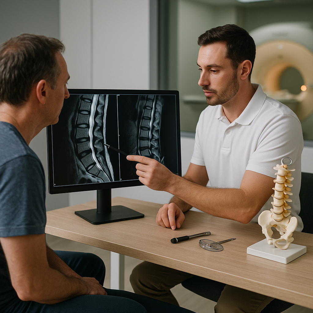
The Role of MRI in Spinal Diagnosis for Chiropractic Patients
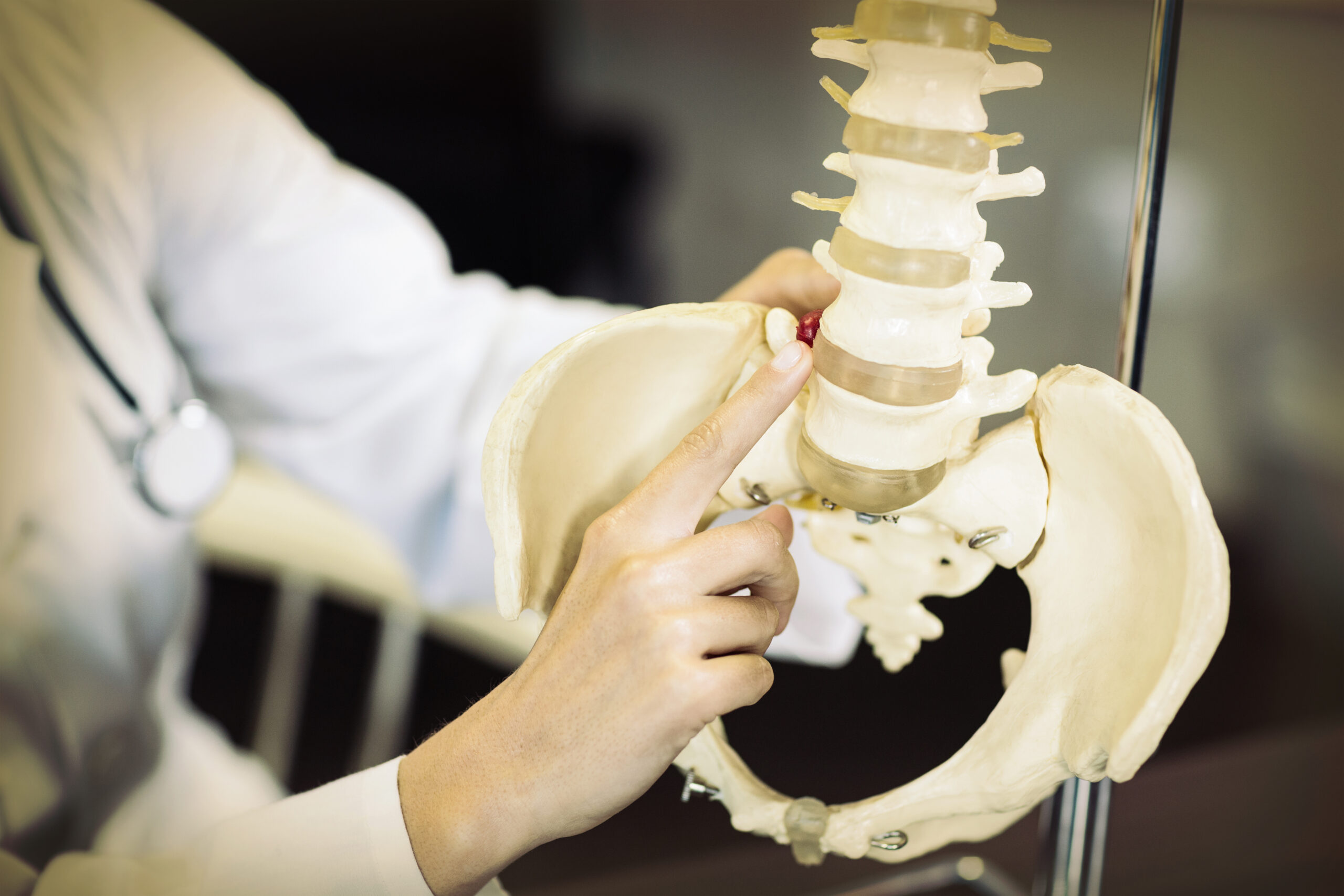
The spine is central to chiropractic practice, and MRI spine diagnosis serves as an invaluable resource for chiropractors managing patients with complex or persistent spinal complaints. The lumbar, thoracic, and cervical regions each present unique diagnostic challenges, and MRI allows for region-specific evaluation with exceptional clarity.
For lumbar conditions, MRI is particularly useful in identifying disc herniations at the L4-L5 and L5-S1 levels — two of the most commonly affected segments in low back pain patients. In the cervical spine, MRI can detect disc protrusions that may be contributing to neck pain, headaches, or upper extremity symptoms. In both cases, the findings obtained from MRI directly influence the course of chiropractic treatment.
By reviewing MRI findings, chiropractors can:
- Determine whether spinal manipulation is safe and appropriate for the patient’s condition
- Identify contraindications to certain chiropractic techniques
- Develop a targeted and individualized treatment plan
- Set realistic expectations for patient outcomes and recovery timelines
- Determine when referral to another healthcare specialist is necessary
Chiropractic Advanced Imaging: Beyond X-Rays
While X-rays remain a fundamental tool in chiropractic diagnostics — particularly for assessing bony alignment, joint space, and vertebral fractures — they fall short when soft tissue pathology is suspected. This is where chiropractic advanced imaging such as MRI becomes indispensable.
The transition from standard radiography to MRI represents a significant leap in diagnostic capability. X-rays offer a two-dimensional view of hard structures, whereas MRI provides multiplanar, high-resolution images of both hard and soft tissues simultaneously. This comprehensive view enables a far more accurate and complete understanding of the patient’s condition.
In addition to MRI, other advanced imaging modalities used in chiropractic diagnostics may include:
- CT scans: Useful for evaluating bony pathology in greater detail than X-rays, particularly in cases of suspected fracture or bone spur formation.
- Diagnostic ultrasound: Increasingly used for real-time soft tissue assessment of tendons, muscles, and peripheral joints.
- Functional MRI (fMRI): Emerging research suggests potential applications for assessing neurological changes associated with chronic musculoskeletal pain.
Despite these options, MRI remains the gold standard for soft tissue visualization and is the most commonly ordered advanced imaging study in chiropractic practice.