Understanding Chiropractic Imaging Tools and Their Role in Patient Care
When you visit a chiropractor for the first time, or when a new health concern arises, your practitioner may recommend a series of diagnostic assessments before beginning any treatment. Among the most valuable of these assessments are diagnostic imaging tools, which allow chiropractors to see beyond the surface and gain a clear, accurate picture of what is happening inside the body. These chiropractic imaging tools are essential for identifying structural abnormalities, ruling out serious conditions, and crafting personalized treatment plans that truly address the root cause of a patient’s discomfort.
Understanding what these tools are, how they work, and why they matter can help patients feel more confident and informed when stepping into a chiropractic clinic. Below, we explore the most commonly used diagnostic tools chiropractors rely on to guide their clinical decision-making.
Why Diagnostic Imaging Matters in Chiropractic Care
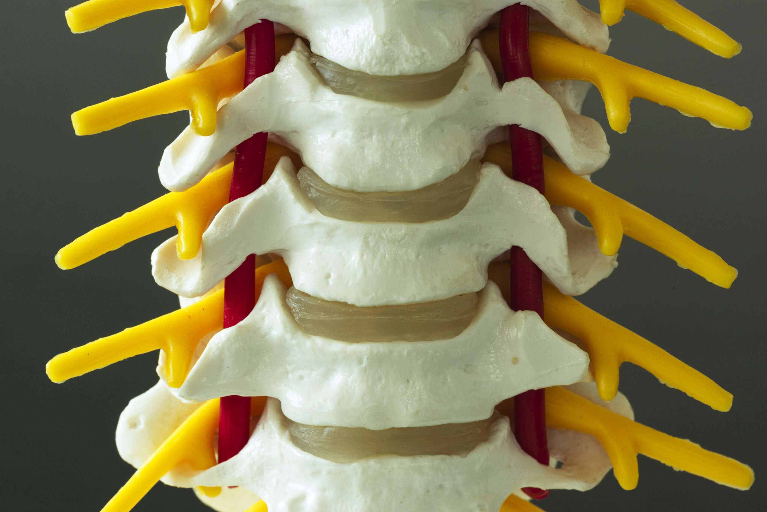
Chiropractic care is built on the understanding that the musculoskeletal system — and particularly the spine — plays a central role in overall health. When the vertebrae, discs, joints, or surrounding soft tissues are compromised, the effects can ripple throughout the body. Without proper imaging, even the most experienced practitioner would be working without a complete picture.
Spine diagnostic equipment gives chiropractors the ability to:
- Identify misalignments, fractures, or degenerative changes in the spine
- Detect conditions that may contraindicate manual manipulation
- Monitor the progress of a patient’s treatment over time
- Differentiate between chiropractic-appropriate conditions and those requiring medical referral
- Provide patients with a visual understanding of their condition
In short, diagnostic imaging bridges the gap between a patient’s reported symptoms and the clinical reality of what is occurring within their body.
X-Ray Imaging: The Foundation of Spine Assessment
Among all chiropractic imaging technology currently in use, X-ray radiography remains one of the most foundational. It has been used in chiropractic practice for well over a century and continues to be one of the first tools a chiropractor may turn to when evaluating a patient’s spinal health.
X-rays produce images of the body’s bony structures by passing a small amount of radiation through the patient’s body. Dense tissues such as bone absorb the radiation and appear white on the resulting image, while softer tissues appear in varying shades of grey. This makes X-rays particularly effective for:
- Detecting vertebral fractures or compression injuries
- Assessing spinal alignment and curvature abnormalities, such as scoliosis or kyphosis
- Identifying signs of degenerative disc disease or osteoarthritis
- Evaluating the spacing between vertebrae, which can indicate disc problems
- Spotting bone spurs, calcifications, or other structural irregularities
Chiropractors are trained to take and interpret spinal X-rays as part of their professional education. Many chiropractic clinics have their own in-house X-ray equipment, while others may refer patients to external imaging centres. Either way, the results provide a critical baseline for care.
It is worth noting that chiropractors follow strict protocols to minimise radiation exposure, taking X-rays only when clinically necessary and using appropriate shielding techniques. The decision to use X-ray imaging is always weighed against the potential clinical benefits for each individual patient.
Magnetic Resonance Imaging (MRI): Visualising Soft Tissue in Detail
While X-rays excel at imaging bone, they have limited capacity to capture soft tissue structures such as intervertebral discs, spinal cord tissue, nerves, ligaments, and muscles. This is where Magnetic Resonance Imaging — commonly known as MRI — becomes an invaluable diagnostic tool chiropractors may request or refer patients for.
MRI uses powerful magnetic fields and radio waves to generate highly detailed cross-sectional images of the body. It produces no ionising radiation, making it a safe option for most patients. The images it creates are remarkable in their clarity, often revealing structures and abnormalities that would remain invisible on an X-ray.
In chiropractic practice, MRI is particularly useful for:
- Diagnosing herniated or bulging intervertebral discs
- Evaluating nerve root compression or spinal stenosis
- Identifying inflammation, oedema, or soft tissue injuries
- Assessing ligament and tendon damage in the spine and surrounding areas
- Ruling out serious pathologies such as tumours, infections, or spinal cord damage
Chiropractors do not typically own or operate MRI machines within their clinics due to the substantial cost and space requirements involved. However, they regularly refer patients to imaging facilities when an MRI is warranted and then incorporate the findings into their treatment planning. A chiropractor who receives an MRI report will analyse it thoroughly to determine whether chiropractic care is appropriate, what adjustments or therapies may help, and whether co-management with a medical specialist is necessary.
Computed Tomography (CT) Scanning: Advanced Bone and Structural Imaging
Computed Tomography, or CT scanning, represents another tier of chiropractic imaging technology. A CT scan uses a series of X-ray images taken from different angles, which a computer then processes to create detailed cross-sectional images — essentially a three-dimensional view of the internal structures.
Additional Resources
- NIH overview of spinal manipulation
- American Chiropractic Association patient resources
- CDC/NIOSH ergonomics resources
- What are the forms required at a first chiropractic visit?
- What are digital health tools used in chiropractic practices?
- What are the questions to ask a chiropractor before treatment?