Understanding Surface Electromyography in Chiropractic Care
If you have ever visited a chiropractor and been asked to stand still while a small device was rolled along your spine, you may have experienced surface electromyography firsthand. Known in clinical practice as sEMG, this non-invasive diagnostic technology has become an increasingly valuable tool in chiropractic assessments. It offers practitioners a window into the electrical activity of the muscles surrounding the spine, helping them build a clearer picture of how the nervous system and musculoskeletal system are functioning together.
Understanding what sEMG chiropractic assessments involve, why they matter, and what the results can reveal is important for any patient seeking a thorough and evidence-informed approach to their spinal health.
What Is Surface Electromyography?
Surface electromyography is a method of measuring the electrical signals produced by muscles when they contract or remain in a state of tension. Every time a muscle fibre activates, it generates a small electrical impulse. By placing sensors on the surface of the skin — rather than inserting needles into the muscle tissue — sEMG technology detects and records these impulses in real time.
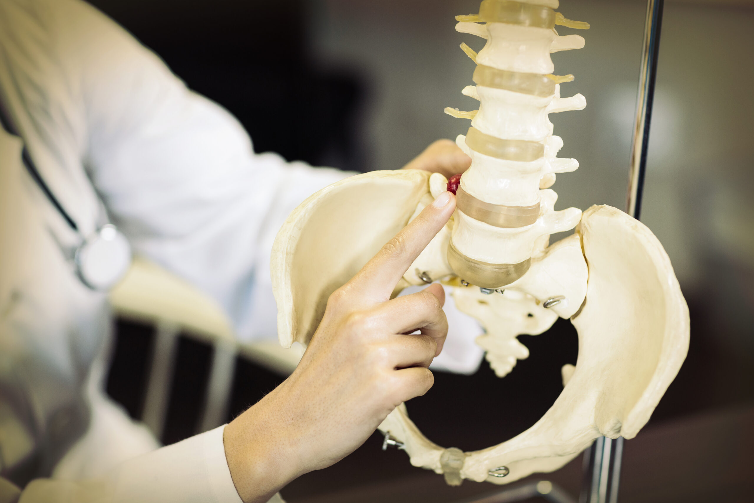
In a chiropractic context, the focus is typically on the paraspinal muscles: the deep and superficial muscle groups that run along either side of the spine. These muscles play a critical role in supporting posture, enabling movement, and protecting the vertebrae. When spinal function is compromised — whether through misalignment, injury, or chronic tension — these muscles often reflect that dysfunction through abnormal electrical activity patterns.
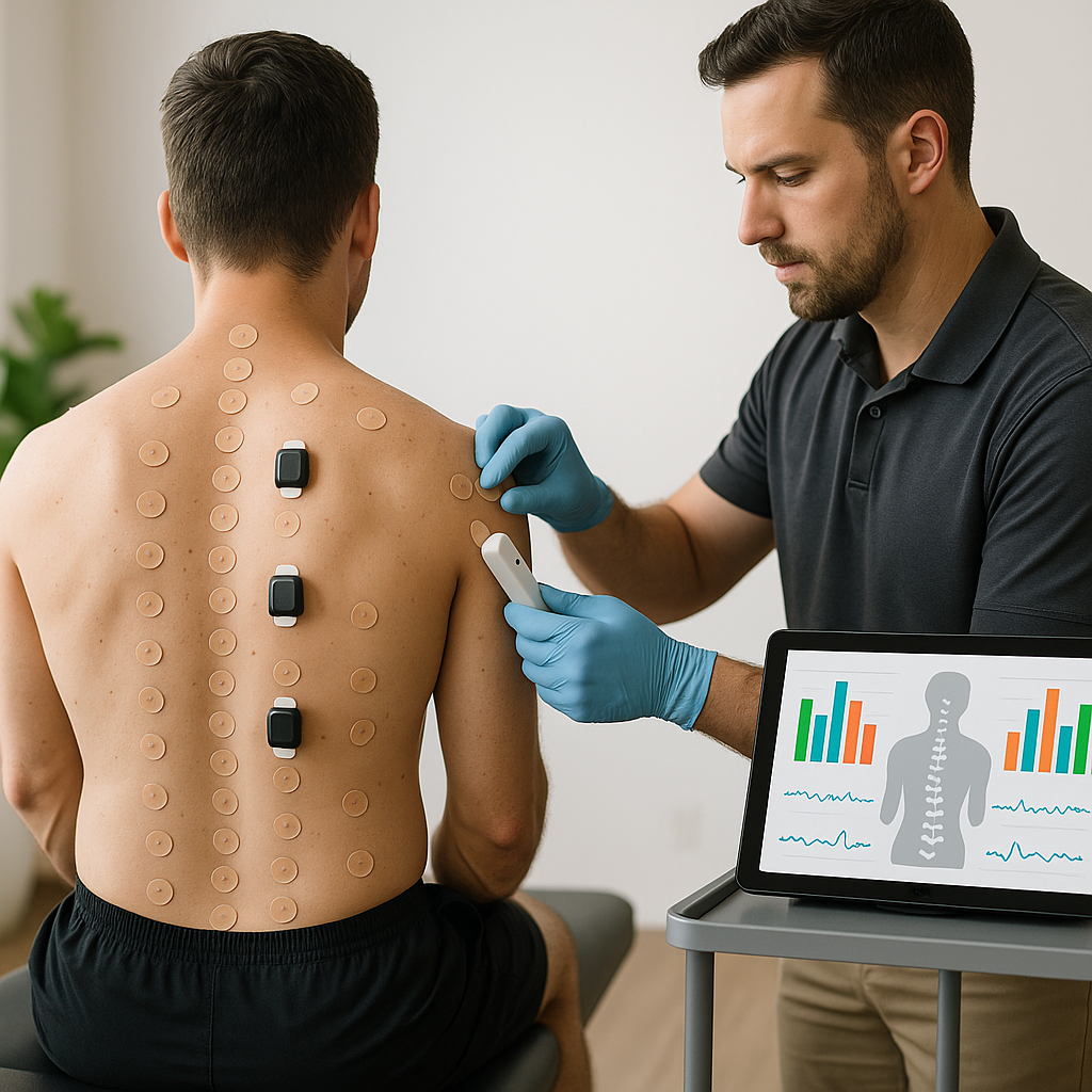
The surface EMG spine assessment is entirely painless. Sensors are either placed directly on specific areas of the back or housed within a scanning device that is gently moved along the spine. The process typically takes only a few minutes and produces a visual readout that the chiropractor can analyse and discuss with the patient.
How Does sEMG Differ from Needle EMG?
It is worth distinguishing surface electromyography from the needle-based electromyography often used in neurological or orthopaedic medical settings. Needle EMG involves inserting fine electrodes directly into muscle tissue to measure activity from within, which can be uncomfortable and carries a small risk of infection or bruising.
Surface EMG, by contrast, uses electrodes placed on the skin’s surface. While this means the signal captures a broader range of muscle activity rather than isolating a single motor unit, it is far more practical for clinical chiropractic use. The surface approach is safe for patients of all ages, requires no preparation, and allows for repeated assessments over time — making it ideal for monitoring progress throughout a course of chiropractic care.
The Role of Paraspinal Muscle Testing in Chiropractic
Paraspinal muscle testing through sEMG provides chiropractors with objective, measurable data about how the muscles along the spine are functioning. This is particularly significant because muscle imbalances and areas of abnormal tension are not always visible during a standard physical examination or even detectable through postural observation alone.
When a vertebra is misaligned or spinal joints are not moving freely, the nervous system often responds by increasing tension in the surrounding muscles. This protective muscular response, sometimes called guarding, can lead to asymmetrical muscle activation patterns where one side of the spine is working significantly harder than the other. Over time, this imbalance can contribute to pain, reduced mobility, and accelerated wear on the spinal structures.
Paraspinal muscle testing allows chiropractors to:
- Identify areas of elevated muscle tension that may not yet be causing noticeable symptoms
- Detect asymmetrical patterns of muscle activity along the spine
- Assess the impact of spinal subluxations on the surrounding musculature
- Establish a baseline measurement against which future assessments can be compared
- Provide patients with a visual representation of their spinal muscle function
This type of objective data strengthens the chiropractor’s ability to design a personalised care plan that addresses the specific needs of the individual rather than relying solely on subjective symptom reports.
What Does a Muscle Activity Scan Involve?
A muscle activity scan using sEMG technology is a straightforward procedure that forms part of a comprehensive chiropractic examination. The patient is typically asked to stand or sit in a relaxed, upright posture while the chiropractor conducts the assessment.
In many modern chiropractic clinics, the sEMG scan is performed using a handheld device that is rolled slowly along the spine from the base of the skull to the sacrum. This device contains sensors that detect the electrical activity of the paraspinal muscles at multiple points along the spinal column. Alternatively, individual electrodes may be placed at specific anatomical landmarks to capture activity at targeted regions.
The data collected is fed into software that generates a graphical display, often presented as a colour-coded chart or bar graph. Areas of high muscle tension are typically represented by larger bars or more intense colours, while regions of normal or balanced activity appear more moderate. This visual output makes it easier for both the chiropractor and the patient to understand the findings quickly and clearly.
The scan itself is completed within a matter of minutes and involves no radiation, no needles, and no discomfort. This makes it a practical and accessible option for routine use in chiropractic practice.
Interpreting sEMG Results
Interpreting the results of an sEMG chiropractic assessment requires clinical expertise and an understanding of normal versus abnormal muscle activity patterns. A well-functioning spine typically shows relatively symmetrical and moderate levels of muscle activity along both sides. Significant deviations from this balance — whether in the form of excessive tension, inhibited activity, or pronounced asymmetry — provide meaningful clinical information.
Elevated readings in a specific spinal region may suggest that the muscles in that area are under chronic stress, possibly due to a vertebral subluxation, joint restriction, or postural imbalance.