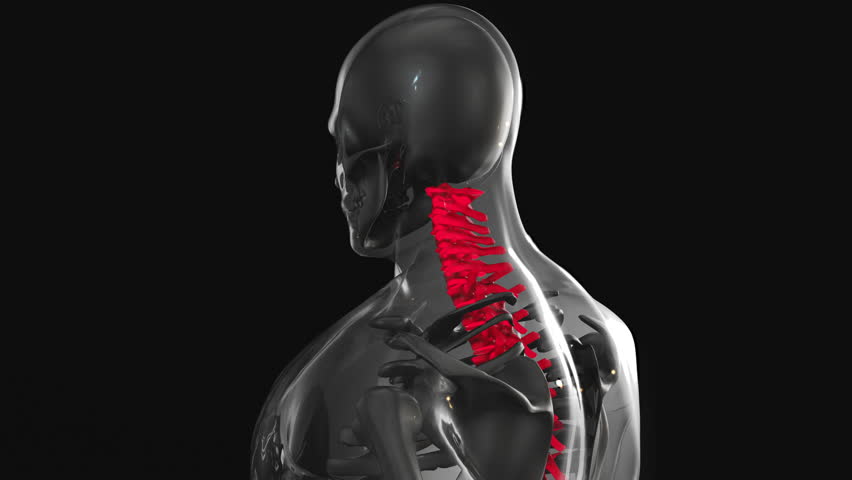
Understanding Motion X-Ray Analysis in Chiropractic Care
When it comes to diagnosing spinal conditions and understanding how the spine behaves during movement, traditional static X-rays only tell part of the story. Motion X-ray analysis in chiropractic — also known as videofluoroscopy spine imaging or dynamic X-ray — offers a far more comprehensive view of spinal health by capturing real-time movement of the vertebrae. This advanced diagnostic tool is transforming how chiropractors assess, diagnose, and treat a wide range of musculoskeletal conditions.
What Is Motion X-Ray Analysis?
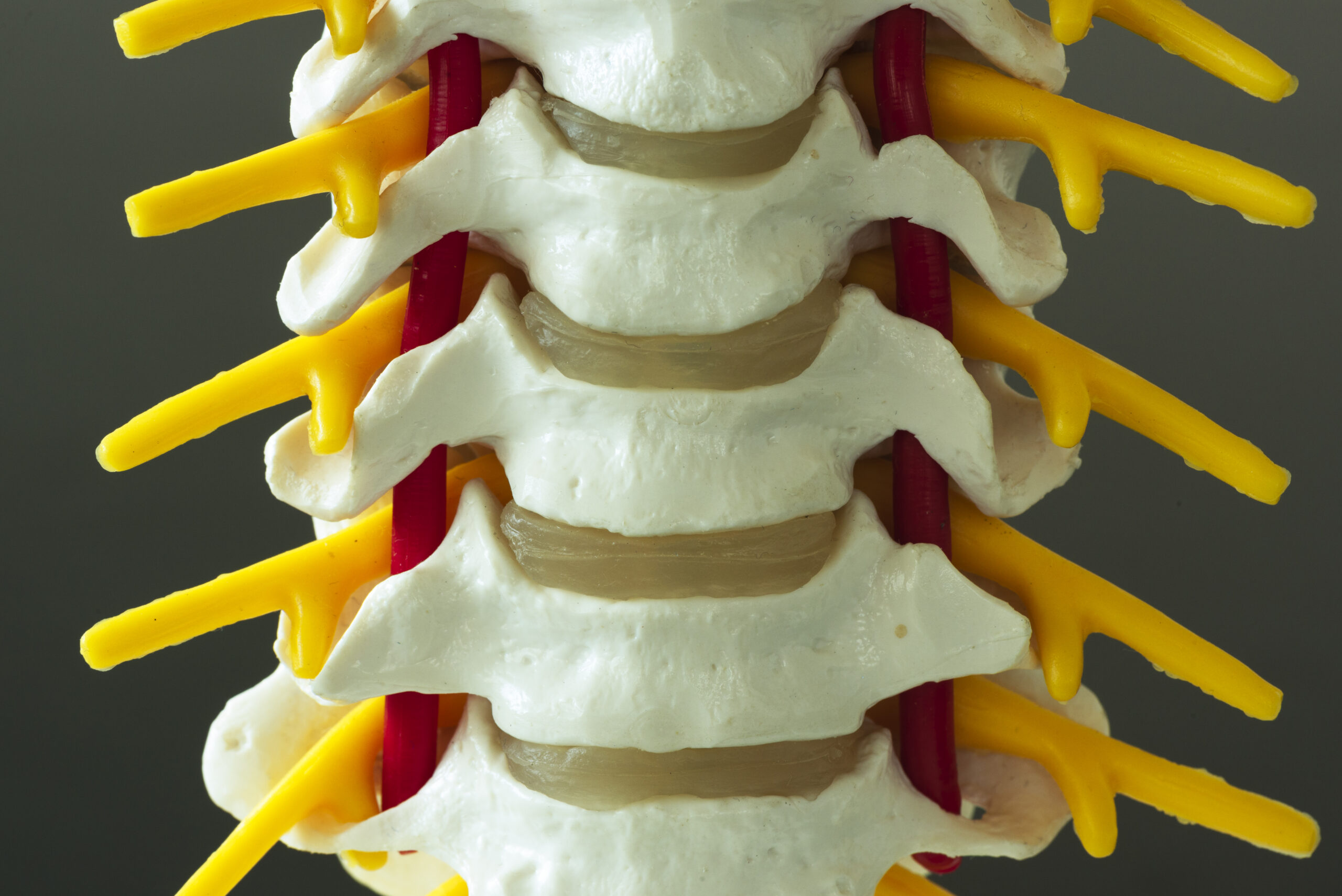
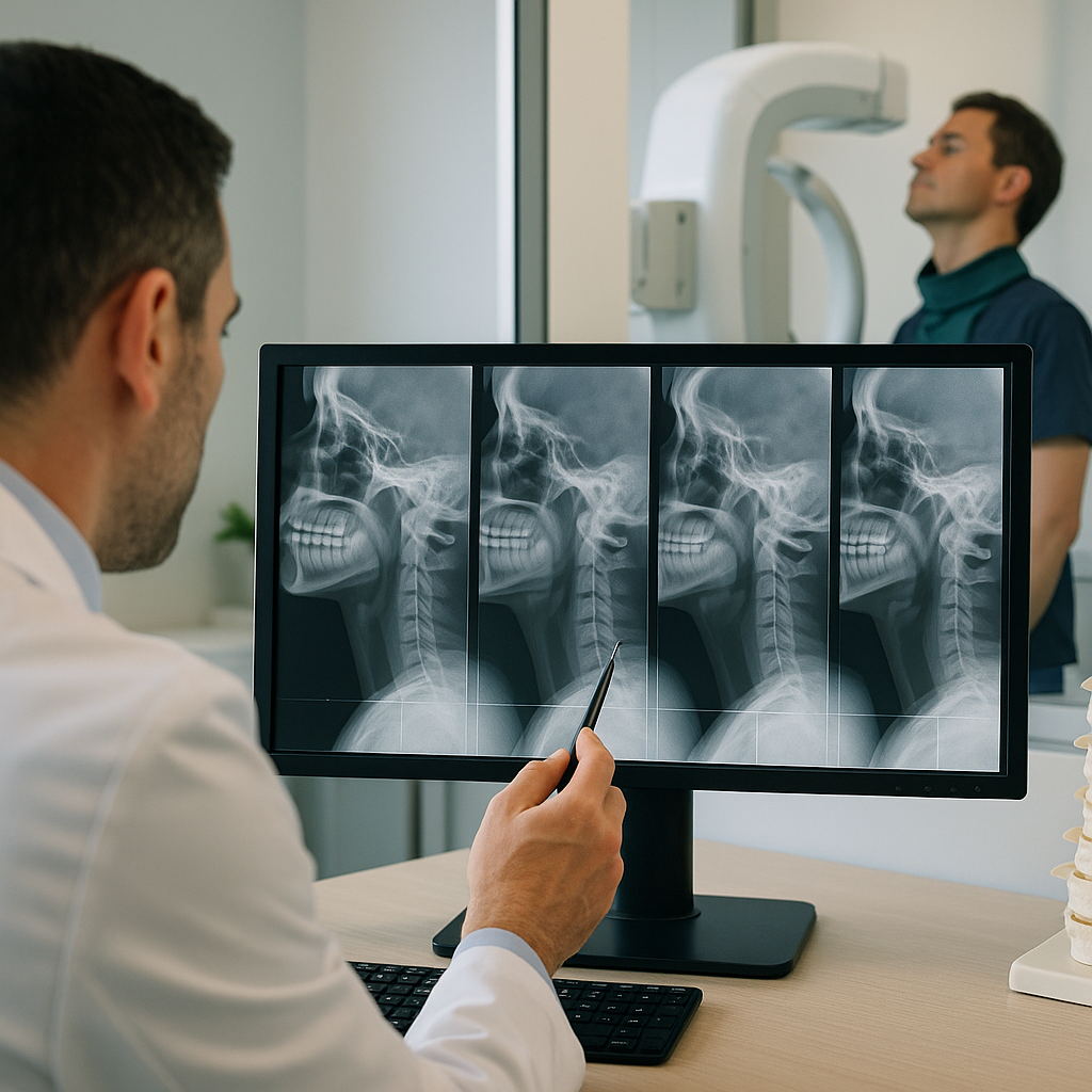
Motion X-ray analysis, clinically referred to as videofluoroscopy or a dynamic spinal motion study, is a diagnostic imaging technique that records continuous X-ray images of the spine while the patient moves. Unlike a standard X-ray that captures a single, still image, motion X-ray chiropractic technology allows practitioners to observe the spine in action — bending forward, leaning backward, rotating, or performing other functional movements.
The procedure uses a fluoroscope, which is a specialized imaging device that produces a live, moving X-ray image, similar in concept to an X-ray video. These images are then reviewed by a qualified chiropractor or radiologist to identify abnormalities in spinal motion that would otherwise remain invisible in static imaging.
How Does the Procedure Work?
The process of undergoing a dynamic X-ray spinal motion study is straightforward and relatively non-invasive. Here is what patients can generally expect:
- Preparation: The patient is positioned in front of the fluoroscopy unit, either standing or seated, depending on the area of the spine being examined.
- Movement Protocol: The chiropractor or technician guides the patient through a series of controlled movements, such as flexion, extension, lateral bending, and rotation.
- Image Capture: The fluoroscope continuously captures X-ray images throughout these movements, creating a dynamic video sequence of the spine in motion.
- Analysis: The recorded footage is reviewed carefully, frame by frame if necessary, to identify areas of abnormal motion, instability, or restricted movement.
The entire procedure typically takes between 15 and 30 minutes, depending on the complexity of the case and the number of spinal regions being evaluated.
Why Is Motion X-Ray Important in Chiropractic?
One of the most significant limitations of conventional diagnostic imaging is that it captures the spine in a fixed position. Many spinal problems, however, only manifest during movement. A vertebra may appear perfectly aligned on a static X-ray but shift abnormally the moment a patient bends or twists. This is precisely where videofluoroscopy spine imaging proves invaluable.
Motion X-ray analysis enables chiropractors to identify:
- Spinal instability: Abnormal shifting or slipping of vertebrae during movement, which may not be visible on standard imaging.
- Hypermobility or hypomobility: Segments of the spine that move too much or too little, both of which can contribute to pain and dysfunction.
- Ligamentous laxity: Looseness in the spinal ligaments that allows excessive motion between vertebral segments.
- Subluxations: Misalignments of the vertebrae that affect nerve function and overall spinal mechanics.
- Post-injury assessment: Particularly useful following car accidents, sports injuries, or falls, where internal damage may not appear on static imaging.
Conditions Diagnosed Through Dynamic X-Ray Spinal Motion Studies
A spinal motion study using videofluoroscopy is particularly beneficial in identifying and evaluating a broad spectrum of conditions. Among the most commonly diagnosed are:
- Cervical instability following whiplash injuries
- Degenerative disc disease and its functional impact
- Spondylolisthesis — the forward slipping of one vertebra over another
- Craniocervical instability (instability at the junction of the skull and upper cervical spine)
- Facet joint dysfunction
- Post-surgical spinal complications
- Chronic neck and back pain with unclear origin
In many cases, patients who have undergone multiple static imaging studies without a clear diagnosis find that a dynamic X-ray finally provides the answers they have been seeking. The ability to observe motion patterns adds an entirely new diagnostic dimension.
Motion X-Ray vs. Traditional Static X-Ray
To fully appreciate the value of motion X-ray chiropractic technology, it is helpful to contrast it directly with conventional static X-ray imaging:
- Static X-Ray: Captures a single moment in time; ideal for identifying fractures, bone density issues, and gross misalignments; cannot assess functional movement.
- Dynamic X-Ray (Videofluoroscopy): Records continuous motion; reveals functional instabilities, movement restrictions, and ligamentous injuries; provides a more complete biomechanical picture.
It is worth noting that both modalities serve important and complementary roles in chiropractic diagnostics. A chiropractor may use static X-rays for initial structural assessment and then recommend a videofluoroscopy spine study when functional abnormalities are suspected or when a patient’s symptoms are not fully explained by standard imaging findings.
Benefits of Motion X-Ray Analysis for Patients
For patients, the advantages of undergoing a dynamic spinal motion study are considerable. The procedure offers:
- Greater diagnostic accuracy: Conditions that are invisible on static imaging become apparent during movement analysis.
Additional Resources