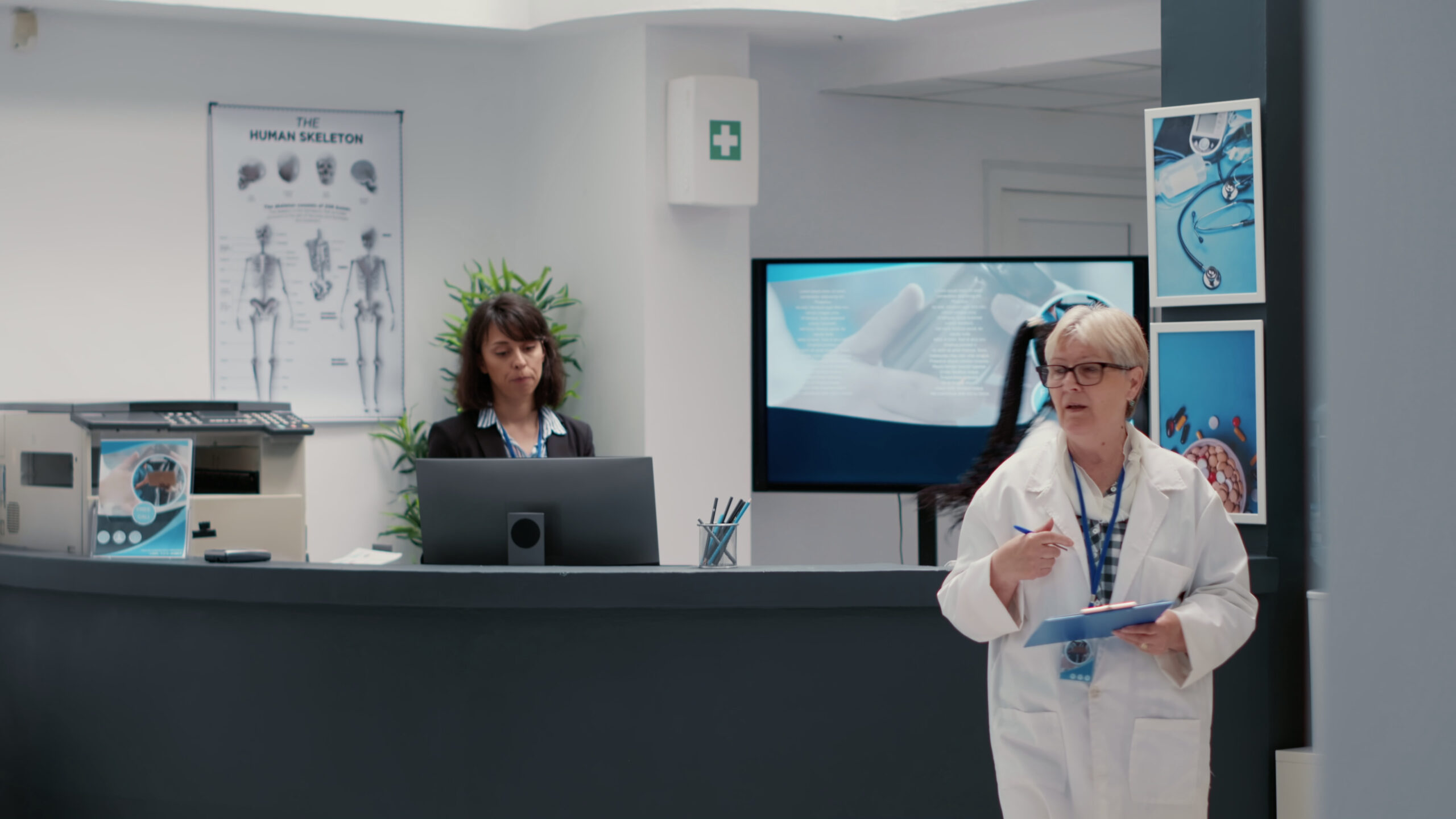
Understanding 3D Spinal Imaging in Chiropractic Care
Modern chiropractic care has evolved significantly over the past decade, and one of the most groundbreaking advancements shaping the field today is 3D spine imaging. Where traditional X-rays once offered only flat, two-dimensional views of the spine, today’s technology allows chiropractors to examine the vertebral column in extraordinary detail — from virtually every angle imaginable. This shift is not merely cosmetic; it represents a fundamental improvement in how spinal conditions are diagnosed, understood, and treated.
Whether you are a patient exploring your treatment options or simply curious about what happens behind the scenes at a modern chiropractic clinic, understanding three-dimensional spinal scanning can help you make more informed decisions about your health. This article breaks down what 3D spinal imaging is, how it works, and why it is becoming an increasingly valued tool in chiropractic practice.
What Is 3D Spinal Imaging?
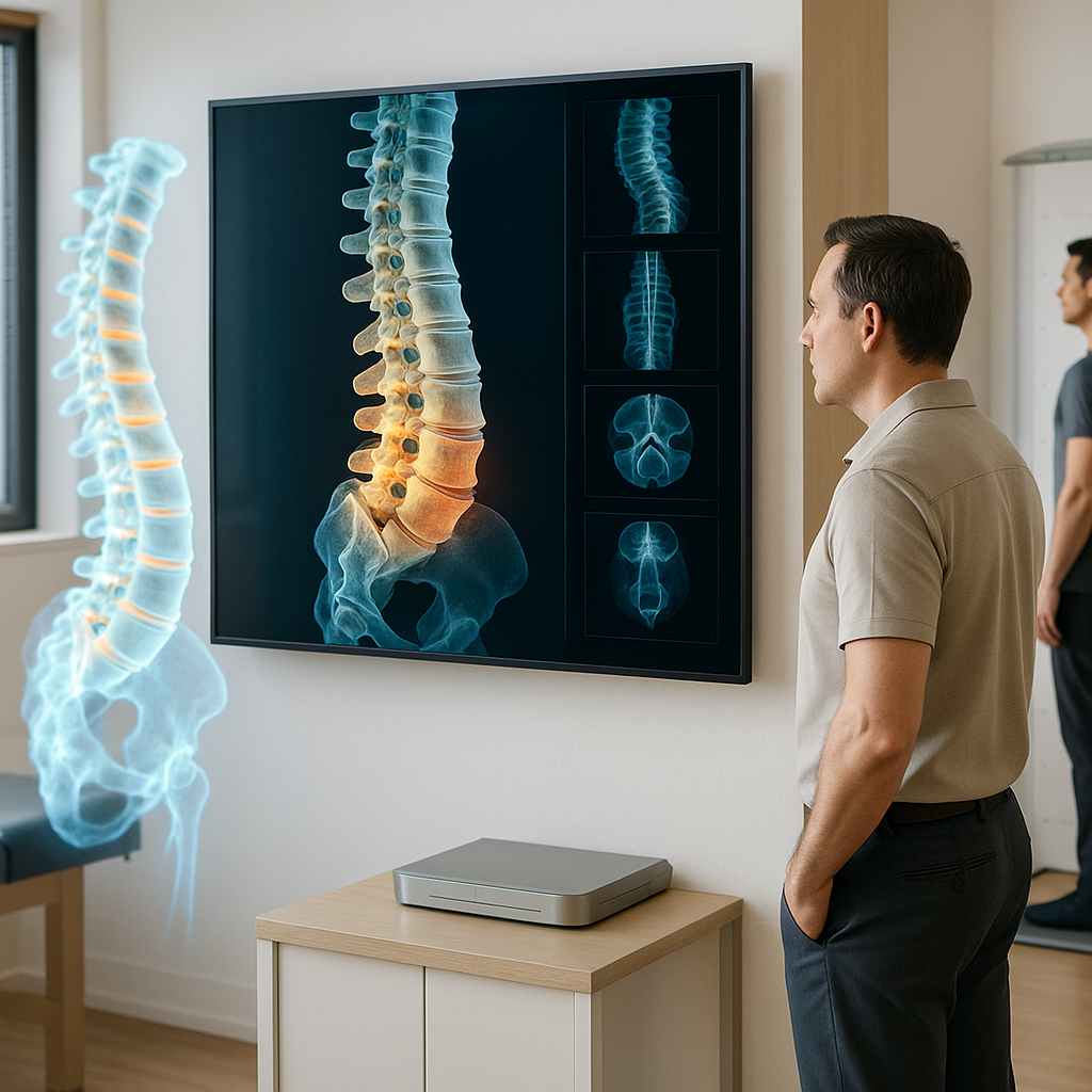
3D spinal imaging refers to advanced diagnostic technology that produces a three-dimensional representation of the spine and surrounding structures. Unlike conventional radiography, which compresses all anatomical layers into a single flat image, 3D imaging captures the spine in volumetric detail, allowing clinicians to rotate, zoom, and examine each segment of the vertebral column independently.
The most widely used technology behind this process is Cone Beam Computed Tomography (CBCT). Originally developed for dental and maxillofacial imaging, CBCT chiropractic applications have expanded considerably as practitioners recognised its superior diagnostic capabilities for musculoskeletal assessment. A CBCT scanner rotates around the patient and captures hundreds of images from different angles in a single sweep. These images are then reconstructed by specialised software into a comprehensive three-dimensional model of the spine.
The result is a highly detailed, interactive digital image that reveals not only the alignment of the vertebrae but also the condition of discs, joints, nerve pathways, and surrounding soft tissue structures — all with remarkable precision.
How Does a Three-Dimensional Spinal Scan Work?
The process of obtaining a three-dimensional spinal scan is straightforward, non-invasive, and typically completed within a matter of minutes. Here is a general overview of what patients can expect:
- Preparation: Patients are asked to remove any metal objects such as jewellery or clothing with metal components, as these can interfere with image quality. No injections, sedation, or special preparation are typically required.
- Positioning: Depending on the type of scanner used, the patient may stand, sit, or lie down. Many CBCT systems used in chiropractic settings allow for upright, weight-bearing scans, which is particularly valuable because it captures the spine under the natural load it experiences during daily life.
- Scanning: The imaging device rotates around the target area, capturing a series of X-ray images from multiple angles within a short time frame — often between 10 and 40 seconds.
- Reconstruction: Advanced software processes the raw image data and reconstructs it into a three-dimensional digital model that the chiropractor can manipulate on screen.
- Analysis: The chiropractor performs a detailed 3D spinal analysis, reviewing the structural relationships between vertebrae, identifying areas of misalignment, degeneration, or abnormality, and developing a treatment plan accordingly.
The radiation dose associated with CBCT imaging is generally lower than that of conventional medical CT scanners, making it a comparatively safer option for diagnostic imaging in a chiropractic setting. Nevertheless, as with all forms of imaging that involve radiation, its use is carefully considered based on clinical necessity.
What Can 3D Spinal Analysis Reveal?
One of the most compelling reasons that 3D spinal analysis is gaining traction in chiropractic practice is the sheer volume and depth of clinical information it provides. Traditional 2D X-rays are valuable tools, but they are inherently limited in their ability to convey the true three-dimensional complexity of the human spine.
A comprehensive 3D spine imaging assessment can identify and evaluate:
- Vertebral alignment and curvature: Conditions such as scoliosis, kyphosis, and lordosis can be assessed with far greater accuracy in three dimensions, helping chiropractors understand the true nature and extent of spinal curvature.
- Intervertebral disc health: While soft tissue visualisation depends on the specific imaging modality, CBCT can identify indirect signs of disc degeneration or herniation through bony landmark assessment.
- Joint and facet condition: Arthritic changes, bone spurs, and joint space narrowing can be clearly identified, contributing to a more accurate differential diagnosis.
- Vertebral subluxations: Chiropractic practice places particular emphasis on identifying vertebral subluxations — subtle misalignments that may interfere with nervous system function. Three-dimensional imaging makes these easier to detect and measure.
- Anatomical variations: Some individuals have congenital or developmental spinal variations that may influence how chiropractic care should be delivered. 3D imaging can reveal these with clarity.
- Post-surgical anatomy: For patients who have previously undergone spinal surgery, 3D imaging helps chiropractors understand the altered anatomy and adjust their approach accordingly.
CBCT Chiropractic Applications: A Closer Look
The integration of CBCT chiropractic technology represents a meaningful step forward in the professionalisation and precision of chiropractic diagnosis. CBCT systems designed specifically for chiropractic use are increasingly available in dedicated clinics and are designed with the musculoskeletal system in mind.
Additional Resources
- NIH overview of spinal manipulation
- American Chiropractic Association patient resources
- CDC/NIOSH ergonomics resources
- What is the difference between traditional and modern chiropractic?
- What are the regions of the spine in chiropractic terms?
- What is the chiropractic approach to breath work and spinal health?