Understanding Sacroiliac Joint Dysfunction and Its Diagnostic Challenges
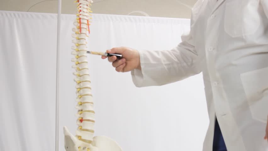
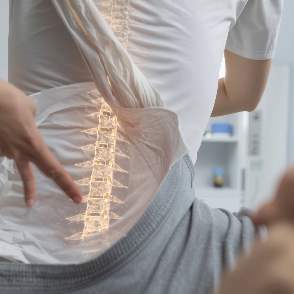
Sacroiliac joint dysfunction (SIJD) occurs when the joints connecting the spine to the pelvis become misaligned or inflamed, leading to chronic pelvic pain. This condition can mimic other issues like sciatica or lower back pain, making accurate diagnosis critical. Patients often experience persistent discomfort that worsens with movement, requiring targeted evaluation. While SIJD is a recognized cause of pelvic pain, its symptoms overlap with other disorders, complicating identification. A thorough diagnostic approach helps differentiate SIJD from similar conditions, ensuring appropriate care. Chiropractors and healthcare providers rely on specific tools and techniques to confirm the diagnosis, as misdiagnosis can lead to ineffective treatments. Understanding the available diagnostic methods empowers patients to engage in informed discussions with their providers about the best evaluation strategies.
Physical Examination Techniques for Identifying SIJD
Manual physical exams are foundational in assessing sacroiliac joint dysfunction. Providers use specific maneuvers, such as the FABER (Flexion, Abduction, External Rotation) test or Gaenslen’s test, to provoke pain and identify joint instability. Palpation of the sacroiliac region helps detect tenderness or asymmetry, while assessing range of motion can reveal restricted movement. These techniques are non-invasive and provide immediate feedback, but their accuracy depends on the provider’s experience. While physical exams are essential, they may not always confirm SIJD definitively. Patients should understand that these evaluations are part of a broader diagnostic process, often combined with imaging or other tools to ensure clarity. A skilled chiropractor can interpret these findings alongside patient history to guide further steps.
Imaging Studies and Their Role in Diagnosis
Imaging studies like X-rays, MRI, and CT scans are commonly used to visualize the sacroiliac joints. X-rays can detect structural abnormalities, such as arthritis or fractures, but may not capture soft tissue issues. MRI offers detailed views of ligaments, discs, and inflammation, making it valuable for identifying SIJD-related changes. CT scans provide high-resolution images of bone structures, aiding in complex cases. However, imaging alone cannot always confirm SIJD, as some patients with joint degeneration may not experience symptoms. Providers often use these tools to rule out other conditions or assess the extent of joint damage. While imaging is a critical component, it is typically combined with clinical findings to ensure a comprehensive diagnosis. Patients should discuss the benefits and limitations of each imaging method with their chiropractor.
Diagnostic Injections and Functional Assessments
Diagnostic injections, such as local anesthetics or corticosteroids, are used to pinpoint the source of pain. A sacroiliac joint block involves injecting a numbing agent into the joint; if pain relief occurs, it supports the SIJD diagnosis. These injections are considered a gold standard for confirmation but are more invasive and carry risks like infection or temporary discomfort. Functional assessments, such as gait analysis or movement pattern evaluations, also help identify SIJD by observing how the body compensates for joint instability. While these methods provide direct evidence, they require specialized training to administer and interpret. Patients should consult with a chiropractor or physician to determine if these tests are appropriate for their situation, as they are often used when other diagnostic approaches are inconclusive.
Emerging Technologies in SIJD Diagnosis
Advancements in technology are expanding diagnostic options for sacroiliac joint dysfunction. Ultrasound, for example, allows real-time visualization of joint structures and can detect inflammation or fluid buildup. Electromyography (EMG) may assess nerve function to rule out neurological causes of pelvic pain. Wearable sensors and motion analysis systems are also being explored to evaluate movement patterns and joint mechanics. While these tools show promise, their use in SIJD diagnosis is still evolving, and clinical evidence varies. Chiropractors may integrate these technologies alongside traditional methods to enhance accuracy. Patients should remain informed about new developments but rely on their provider’s guidance to determine which tools are most suitable for their case.
Combining Diagnostic Approaches for Accurate Results
A comprehensive diagnosis of sacroiliac joint dysfunction typically involves combining multiple methods. Physical exams, imaging, and injections each offer unique insights, and their integration improves accuracy. For example, a positive physical exam finding paired with MRI evidence of inflammation strengthens the SIJD diagnosis. Providers may also consider patient history, symptom patterns, and response to conservative treatments. This multi-faceted approach reduces the risk of misdiagnosis and ensures targeted care. Patients should work closely with their chiropractor to understand how these tools are used in their evaluation. Open communication about symptoms and concerns helps providers tailor the diagnostic process, leading to more effective treatment plans. Ultimately, a collaborative effort between patient and provider is key to addressing chronic pelvic pain effectively.