Understanding the Conditions: Piriformis Syndrome vs. Sciatica
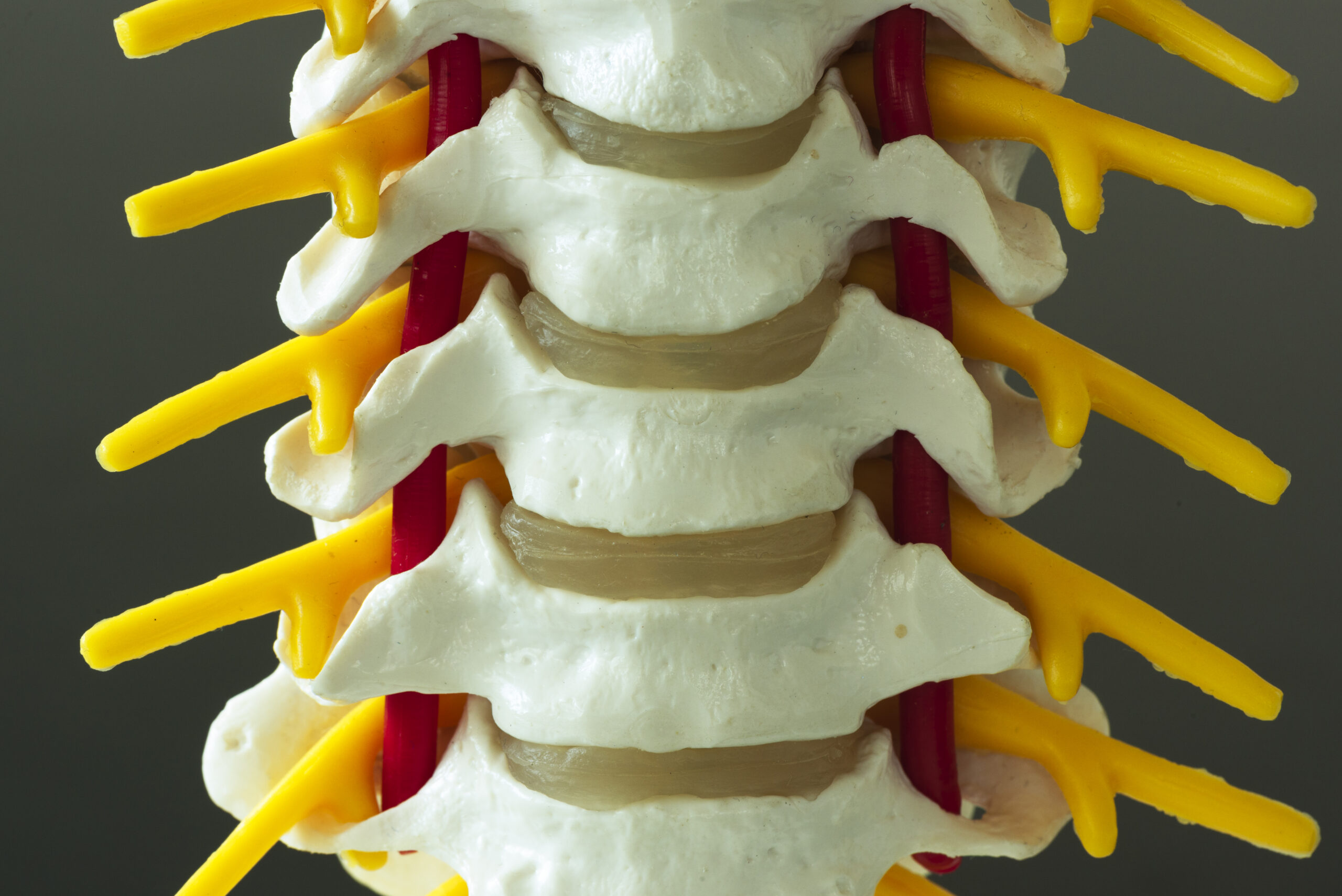
Piriformis syndrome and sciatica both involve nerve-related pain in the lower back and legs but differ in their underlying causes and symptom patterns. Piriformis syndrome occurs when the piriformis muscle, located in the buttocks, compresses the sciatic nerve, leading to pain, tingling, or numbness that radiates from the lower back down the leg. Sciatica, on the other hand, refers to pain along the sciatic nerve pathway caused by compression or irritation of the nerve roots in the spine, often due to a herniated disc or spinal stenosis. While both conditions share similar symptoms, their origins and diagnostic approaches vary. Chiropractors must recognize these distinctions to provide accurate assessments and effective care. Understanding the anatomical differences and typical patient presentations is critical for differentiating between the two during a physical exam.
Patient History and Symptom Analysis
A thorough patient history is essential for distinguishing piriformis syndrome from sciatica. Patients with piriformis syndrome often report pain that worsens with prolonged sitting, walking, or activities that engage the piriformis muscle, such as climbing stairs. The pain may be localized to the buttocks and radiate down the back of the leg but typically does not extend below the knee. In contrast, sciatica pain usually originates from the lower back and travels along the sciatic nerve, often reaching the foot. Patients may describe a sharp, burning, or shooting pain, accompanied by numbness or weakness. Chiropractors should ask about the onset of symptoms, aggravating factors, and any prior injuries or conditions. This information helps narrow down potential causes and guides the physical examination process.
Physical Examination Techniques for Differentiation
During a physical exam, chiropractors use specific maneuvers to identify the source of nerve-related pain. The Freiberg’s test, which involves flexing the hip and knee while the patient lies on their side, can provoke pain in piriformis syndrome. The FAIR (Flexion, Adduction, and Internal Rotation) test, where the examiner applies resistance while the patient’s leg is moved into a specific position, is another indicator of piriformis involvement. For sciatica, the straight leg raise test is commonly used; pain radiating below the knee during this maneuver suggests nerve root compression. Chiropractors also assess muscle strength, reflexes, and range of motion to detect abnormalities. These techniques, combined with patient feedback, help determine whether the issue stems from the piriformis muscle or spinal structures.
Differential Diagnosis Considerations
Chiropractors must consider other conditions that may mimic piriformis syndrome or sciatica, such as sacroiliac joint dysfunction, lumbar radiculopathy, or hip pathology. For example, sacroiliac joint issues often cause pain in the lower back and buttocks that worsens with standing or walking, while hip disorders may present with groin or thigh pain. A comprehensive exam includes evaluating the lumbar spine, pelvis, and surrounding musculature to rule out these possibilities. Imaging studies, such as X-rays or MRI, may be necessary if the diagnosis remains unclear. However, the physical exam remains the cornerstone of initial assessment. By systematically addressing each potential cause, chiropractors can improve diagnostic accuracy and tailor treatment plans to the patient’s needs.
Imaging and Diagnostic Tools
While physical exams provide critical insights, imaging and diagnostic tools can supplement the assessment of piriformis syndrome and sciatica. X-rays may reveal spinal abnormalities, such as herniated discs or degenerative changes, that contribute to sciatica. MRI scans offer detailed views of soft tissues, helping identify piriformis muscle hypertrophy or sciatic nerve compression. Electromyography (EMG) can assess nerve function and confirm nerve root irritation. However, these tools are not always necessary for initial diagnosis and are often used when symptoms persist or when other conditions are suspected. Chiropractors should interpret imaging results in conjunction with clinical findings, ensuring a holistic approach to patient care. The goal is to use technology to support, not replace, the physical examination process.
Clinical Decision-Making and Follow-Up
After gathering patient history, performing physical exams, and reviewing diagnostic findings, chiropractors must synthesize the information to make an informed diagnosis. Treatment plans vary depending on the condition, with piriformis syndrome often involving soft tissue therapy, stretching, and posture correction, while sciatica may require spinal adjustments, decompression, or referral for further intervention. Follow-up appointments are crucial to monitor progress and adjust care as needed. Chiropractors should educate patients on self-management strategies, such as avoiding aggravating activities and incorporating therapeutic exercises. Collaboration with other healthcare providers may also be necessary for complex cases. By maintaining a patient-centered approach, chiropractors can enhance outcomes and ensure long-term relief for individuals experiencing nerve-related pain.